Click Here for More Images from iStock- 15% off with coupon 15FREEIMAGES 
Motor Neuron cells under the microscope
plant Zea Stem C.S. under light mircoscope with white background
Newport oregon
corn stem micrograph with dye
Illustration of animal cell anatomy
Photomicrograph of flatworm,
Epidermis. Electron microscope micrograph showing a keratinocyte of spinous layer. The epithelial cell has a polygonal shape, central nucleus with nucleolus, cytoplasm full of keratin filament bundles, and numerous dark desmosomes crossing the intercellular spaces.
Radish root, cross section under the light microscope. Traverse section through the root of the Raphanus sativus plant. Light micrograph at 8X magnification. Isolated, on white Background.
Couple of two jellyfishes flowing under the sea
Leiden leaf c.s. under microscope
Natural agate surfaces, suitable for backgrounds and wallpapers.
Common jellyfish, Aurelia aurita, Cannonball jellyfish, Stomolophus meleagris, Purple-striped jellyfish, Chrysaora colorata and Disc jellyfish, Sanderia malayensis, against white background
A jellyfish glows from within creating a surreal scene. The soft glow hides the danger associated with these sea creatures.
Mediterranean jelly or fried egg jellyfish or Mediterranean jellyfish (Cotylorhiza tuberculata), isolated on white background. Easy photoshop with the original underwater photo.
plant rape flower cross section under light mircoscope with white background
Underwater jellyfish photos cut out on a white background
A peek inside a jellyfish, floating underwater
ascaris megalocephala cross section under the microscope showing ovaries - optical microscope x200 magnification
A saltwater jellyfish.
3d rendering of Giardia, is a microscopic parasite that lives in the intestines. The parasite can cause a bowel infection called
Diatom or yellow-green alga. Each diatom is composed of one cell and is mobile. San Francisco Bay, California, USA. Live specimen. Wet mount, 40X objective, transmitted brightfield illumination. Note - motion blur of live specimen, very shallow depth of field, chromatic aberration and uneven focus are inherent in light microscopy.
a Close Up Of Jellyfish Swimming In Sea
Blastomycosis is a fungal infection caused by inhaling Blastomyces dermatitidis spores. If it involves only the lungs, it is called pulmonary blastomycosis. Only about half of people with the disease have symptoms, which can include fever, cough, night sweats, muscle pains, weight loss, chest pain, and feeling tired.
The study of plant tissues under the microscope in the laboratory.
Oviduct T.S. under light microscope with white background
Cells consist of a protoplasm enclosed within a membrane, which contains many biomolecules such as proteins and nucleic acids.
Close-up shooting of a green agate stone mineral surface isolated on white background. Its a macro shot shows nature beautiful details with back lit. Agate is a kind of natural gemstone. Abstract pattern. Good for Wallpaper texture background. Turquoise, blue
realistic 3d render of jellyfishes
Aurelia aurita (also called the common jellyfish, moon jellyfish, moon jelly or saucer jelly) is a species of the family Ulmaridae. The jellyfish is almost entirely translucent. Monterey Bay, California.
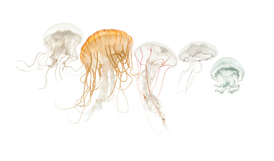
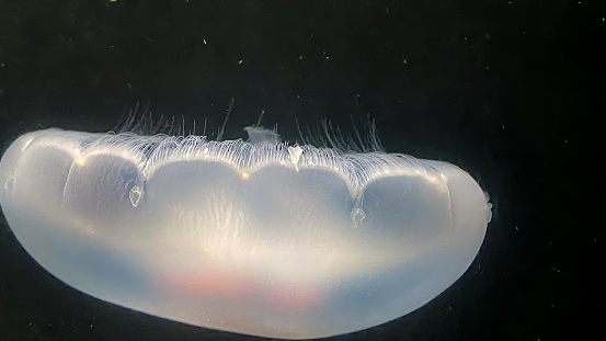
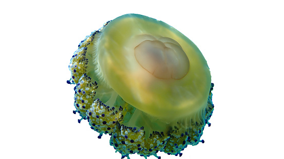
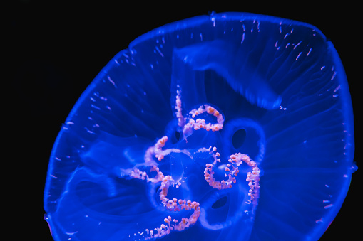
Free Images: "bestof:science diagram cross drawing fish body section parts system biology jelly labelled labeled jellyfish part"
fossil-petrification-fish-skeleton-201044.jpg
Protecting_Tranquilized_Bea.jpg
hand-magnifying-glass-discovery-1701969.jpg
science-diagram-cross-drawing-fish-41526.svg
Jellyfish cross section numbered.svg
school-education-science-diagram-40847.svg
skeleton-human-diagram-labelled-41513.svg
brain-diagram-medical-biology-40356.svg
Cross section jellyfish en.svg
Little Boy internal diagram.svg
Cross section jellyfish ru.svg
diagram-fruit-labelled-labeled-41532.svg
Flag of Croatia for discussion.svg
red-science-diagram-cell-neuron-41524.svg
lungs-organ-biology-respiratory-39980.svg
stomach-anatomy-human-body-biology-310730.svg
skeletal-labeled-worksheet-skeleton-40500.svg
cerebellum-cerebrum-brain-science-40355.svg
eye-diagram-eyeball-body-pupil-39998.svg
Design_Analysis_of_the_P-38_Lightning.png
Globe valve diagram.svg
Axial skeleton diagram blank.svg
Little Boy internal diagram (no labels).svg
Endomembrane system diagram en (edit).svg
Examples of Airfoils.svg
Plant cell structure-en.svg
Plant cell structure svg labels.svg
Plant cell structure svg 2.svg
Design_Analysis_of_the_B-24J_Liberator.png
Design_Analysis_of_the_Boeing_B-17G_Flying_Fortress.png
red-science-diagram-cell-biology-41576.svg
seed-biology-diagram-science-41533.svg
Design_Analysis_of_the_Lockheed_PV-1_Ventura.png
skeleton-bones-human-science-294251.svg
digestive-system-human-digestion-41529.svg
human-body-circulatory-system-41635.svg
circulatory-system-labels-biology-41523.svg
lungs-diagram-air-system-39983.svg
lungs-human-diagram-respiratory-39981.svg
lungs-organ-human-diagram-medicine-37824.svg
Tooth Section numbered.svg
Intestine.svg
Pioneer plaque man as diagram template.svg
Tree secondary components diagram.svg
human-heart-blood-flow-1700453.svg
Pioneer plaque man upper body as diagram template.svg
Incandescent light bulb (no labels).svg
Pioneer plaque man upper body as diagram template (alternative).svg
planet-chalk-drawing-celestial-body-381127.jpg
chalk-drawing-celestial-body-381126.jpg
Submarine cable cross-section 3D plain.svg
Axial skeleton diagram.svg
Gray5.svg
Gray1167.svg
Gray1216.svg
Gray5-ja.svg
Gray1167_fi.svg
Penis_mk.svg
hand-finger-show-touch-success-577356.png
hand-finger-show-touch-success-577355.png
Gray1167 es.svg
Mature flower diagram ar.svg
Gray5_de.svg
Gray5_nl.svg
Gray5_-_es.svg
Mature flower diagram.svg
Mature flower diagram TR.svg
Scheme female reproductive system-hu.svg
Gray1167_pl.svg
Gray814_pl.svg
Table_of_Affinity_-_Google_Art_Project.jpg
Scheme female reproductive system-hu (1).svg
Incandescent light bulb.svg
CooperRiverBridge.svg
Animal mitochondrion diagram en.svg
Animal mitochondrion diagram bn.svg
Animal mitochondrion diagram en (edit).svg
British Isles Euler diagram 15.svg
Design_Analysis_of_the_F6F-5_Hellcat.png
Surface projections of the organs of the trunk.png
Longcase_clock_movement.png
Blood-vascular_system_of_the_frog.jpg
Sexualidadvaron.PNG
Diagrams_of_blood_circulation_in_mamamal,_bird,_reptile_and_fish.jpg
Cross_section_of_lock_from_the_Panama_Canal.jpg
Verge_Escapement_Labelled.png
Swedish Atomic Bomb.png
Gray974.png
Gray527.png
The_Quarterly_journal_of_the_Geological_Society_of_London_(12512113463).jpg
Floyd rose pro parts.jpg
The_Quarterly_journal_of_the_Geological_Society_of_London_(12645922603).jpg
Projected carbon dioxide emissions and atmospheric concentrations over the 21st century for reference and mitigation scenarios.png
Stetefeldt roasting furnace.png
An_Exact_Plan_and_Section_of_Old_Sarum.jpg
Design_Analysis_of_the_Vought_F4U-1D_Corsair.jpg
Illu_repdt_male.jpg
FMIB_43119_Sectional_elevation_and_cross_section_improved_call_system_fishway-_Approved_by_United_States_Fish_Commission.jpeg
IGBT Cross Section.jpg
Illu repdt male gl.jpg
Terms of Use   Search of the Day