Click Here for More Images from iStock- 15% off with coupon 15FREEIMAGES 
GABA B receptors  are G protein-coupled receptors, also called metabotropic receptors.  Binding of  an agonist (baclofen, red) leads to a G-protein coupled C-AMP signaling pathway. Source: PDB entries 7eb2, 6r3q,.
The three-dimensional structure of proteins is represented in a variety of conformations. This image is based on the ribbon structure of a protein. It is suitable for biology notebooks and book covers.\nThis image has not been created by AI.
3D cartoon and Gaussian surface models, PDB 6vg8, white background
A ball and stick + ribbons model of beta-secretase - a protein that is important in forming healthy peripheral nerve cells, but is also involved in the onset of Alzheimer's disease.
Nucleosome structure. Structure of an oligonucleosome, showing the packaging of DNA in chromosomes. 3D illustration.
Molecular Model of Human Insulin with disulfide bonds shown.
Covid-19 Coronavirus Variants Omicron Delta Gamma Beta Alpha Pandemic Virus Disease Mutation
DNA mutation, a mutation is a change in a DNA sequence, mutations can result from DNA copying mistakes made during cell division.
The glucagon like peptide-1 receptor (GLPr) is a G proteincoupled receptor (GPCR) that mediates the action of GLP-1, a peptide hormone. The activated receptor  has a strong effect  on the management of type 2 diabetes mellitus and obesity, including glucose homeostasis and regulation of gastric motility and food intake.
DNA Helix - 3d rendered image. Genetic research concept. Complex spiral structure illustration. Medical, science, genetic biotechnology, gene cell, chromosome idea. DNA strands on dark background . Science nanotechnology.
3D cartoon and Gaussian surface models, chain id color scheme, PDB 3omv, white background
Artificial Intelligence digital concept
Digital medical illustration: Microscopic view of human DNA (PDB-1BNA). Based on actual molecular data.
dna close-up 3D
A ball and stick + ribbons model of human Interleukin 2 or IL-2. Interleukin is a hormone which acts as a signalling molecule for the immune system.
EBP Evidence based practice Concept. Chart with keywords and icons on white desk with stationery.
After dopamine binding, the dopamine receptor changes his shape and binds the inactive G protein.The G protein expels its GDP molecule replacing it by GTP. and falls into two pieces. The Galpha subunit binds to and activates the enzyme adenylyl cyclase, which produces then cyclic AMP spreading a signal through the cell.
DNA Helix - 3d rendered image. Genetic research concept. Complex spiral structure illustration. Medical, science, genetic biotechnology, gene cell, chromosome idea. DNA strands on dark background . Science nanotechnology.
3D cartoon and Gaussian surface models, PDB 7las, chain id color scheme, white background
Inside protein molecule
Tetradecylthioacetic acid molecule. 3D rendering. Atoms are represented as spheres with conventional color coding.
Micrograph of amoeba.  Live specimen. Wet mount, 40X objective, transmitted brightfield illumination. Note - motion blur of live animal, very shallow depth of field, chromatic aberration and uneven focus are inherent in light microscopy.
Isolated Poly Protein G with Detection Antibodies to enhance Immunoassays sensitivity 3d rendering
molecular structure digital concept
The glucagon like peptide-1 receptor (GLPr) is a G proteincoupled receptor (GPCR) that mediates the action of GLP-1, a peptide hormone. The activated receptor  has a strong effect  on the management of type 2 diabetes mellitus and obesity, including glucose homeostasis and regulation of gastric motility and food intake.
DNA and biotechnology concept
Resistin also known as adipose tissue-specific secretory factor (ADSF) or C/EBP-epsilon-regulated myeloid-specific secreted cysteine-rich protein (XCP1) is a cysteine-rich peptide hormone derived from adipose tissue that in humans is encoded by the RETN gene. 3D cartoon and Gaussian surface models, chain id color scheme, based on PDB 1rfx, white background
3d render of abstract connection background
Chain of amino acid or bio molecules called protein - 3d illustration
5G Mobile Network Data Technology, Global Communication, Speed
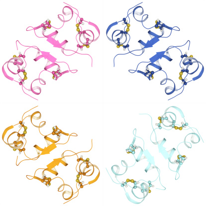
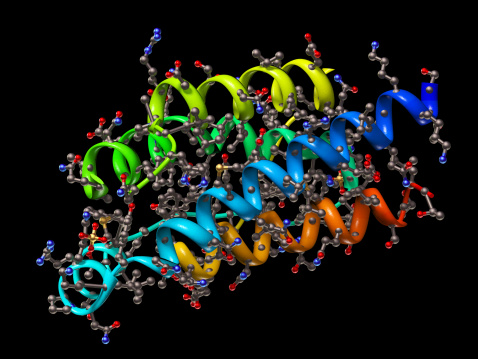
Free Images: "bestof:PDB 2ret EBI.png Alpha and beta proteins a+b Pili subunits Pili subunits EpsJ-like Type II secretory pathway component EpsJ Alpha and beta proteins a+b Pili"
PainleveTypeIII.svg
Comparison of properly and improperly scaled picture graph.svg
Ferrier carbocyclization.svg
Mm'-Type Low-pass Transfer Function (1 Half-section).svg
2ph1 pdb gallery nucleotide binding-protein-2 (MinD homolog).svg
Reserpine 200.svg
4-hydroxy-3-(3-oxo-1-phenylbutyl)-2H-chromen-2-one 200.svg
@marcelosilksantos_-_vector-estampa-camiseta-vetor-em-corel-draw-vetorizao-imagens-vetorizadas-silk-screen-serigrafia-branco-e-preto-FORMANDOS-_TERCEIRÃO-_COREL-_(119).jpg
Beta alpha approx.svg
Alpha beta.svg
17-alpha- vs 17-beta-estradiol.svg
Resolve triangle with c alpha beta.svg
Maltulose.svg
Cysten a-b eliminierung.svg
Gulose.svg
Ergoloid skeletal.svg
Ergotoxine skeletal.svg
1,2-, 1,3- & 1,4-Tropolone.svg
Ferrier carbocyclization 2.svg
Glucose numbering.svg
Mannose structure.svg
Rete.svg
Alpha-methylstyrol.svg
SilylEnolEtherFromHydrosilylation.svg
Constant-k Filter Transmission Parameters.svg
PKARII.svg
Avoparcins.svg
Imperialine.svg
Alpha-cedrene skeletal.svg
Beta-cedrene skeletal.svg
Complement pathway.svg
Zelula prokarioto tipikoa.svg
NFA regexp concat.svg
NFA regexp sum.svg
Equilateral pentagons plot.svg
Prokaryote cell-ro.svg
Ketocarboxylic Acids General Formulae V.1.svg
Tabtoxin.svg
Tabtoxinine beta-lactam.svg
(S)-Acetolactic acid.svg
Acetolactic acid.svg
Average prokaryote cell- nl.svg
Picture Graph.svg
Fixed-points theory and variance minimization (dynamic vibration absorber) (SVG).svg
Triangle-law-of-sines.svg
Triangle-law-of-sines-2.svg
William_Louis_Sonntag_-_Landscape.jpg
Arotinolol.svg
UnitCell.svg
Equilateral pentagon triangulated.SVG
Constant-k Filter Gain with Multiple Half-Sections.svg
Integrality of root systems.svg
Raumzeigerdarstellung.svg
Chaucer Astrolabe (Rete).svg
Ferrier carbocyclization mech.svg
Okuns law with confidence bands.svg
Amnioacids amine gpt position.svg
Improperly scaled picture graph.svg
Proteinoxidation 03.svg
Error centring.svg
Dirichlet process draws.svg
S-R-processes-atomic-mass-201-to-210.svg
Landscape_by_William_Louis_Sonntag,_after_1857,_oil_on_canvas_-_Chazen_Museum_of_Art_-_DSC02158.JPG
Equilateral pentagon.SVG
M-Derived Low-pass Transfer Function (1 Half-section).svg
Sinussatz.svg
2-bromo-1-phenylethanone 200.svg
Average prokaryote cell- ru.svg
Average prokaryote cell- uk.svg
星象图.svg
Stability region for BDF1.svg
(S)-2-aminobutanoic acid 200.svg
Hepatitis B virus v2.svg
Kt arbre epaule torsion.svg
Kt arbre epaule traction.svg
Kt arbre epaule flexion.svg
Triangle-right-abchpq.svg
Conjugate gradient illustration.svg
2-butoxy-N-(2-(diethylamino)ethyl)quinoline-4-carboxamide 200.svg
Civil Air Ensign of Australia.svg
P450cycle.svg
PDB 2ret EBI.png
PDB 3ci0 EBI.png
PDB 1x6z EBI.jpg
PDB 1x6x EBI.jpg
PDB 1x6q EBI.jpg
PDB 1x6y EBI.jpg
PDB 1x6p EBI.jpg
PDB 1x6r EBI.jpg
PDB 1dzo EBI.jpg
PDB 1oqw EBI.jpg
PDB 1q5f EBI.jpg
PDB 2pil EBI.jpg
PDB 1ay2 EBI.jpg
PDB 1qve EBI.jpg
PDB 1rg0 EBI.jpg
PDB 1hpw EBI.jpg
PDB 1oqv EBI.jpg
PDB 1t92 EBI.jpg
PDB 2gr8 EBI.jpg
Terms of Use   Search of the Day