Click Here for More Images from iStock- 15% off with coupon 15FREEIMAGES 
An MRI of the whole spine image is a comprehensive visual representation produced through Magnetic Resonance Imaging, providing detailed insights into the entire spinal structure.
MRI of spinal column
Osteoporosis is a systemic skeletal disorder characterized by increased fragility and deterioration of the bone tissue. Osteoporosis is caused by an imbalance of osteoblast and osteoclast activity.
Cervical Vertebrae Bridge X-ray Film
X-ray image of a normal human spine.
Abdominal X-Ray Techniques
Computational tomography image of the abdominal and pelvic regions.
CT SCAN of Chest and Abdomen Coronal and sagittal view with injection contrast media for diagnosis chest and abdomen disease.
X-ray of female hips, pelvis and spine
Backbone and joint diagnostics. Lumbar and skeleton test.
X-ray of the human mouth showing dental
set of 6 sagittal MRI scans of neck area of 34 years old caucasian male with herniaset of MRI scans of neck area of caucasian male with bilateral paramedial, with a left accent, extrusion of the C6-C7 segment with left-sided C7 radiculopathy, moderate paresis of the triceps of the left hand.
Human neck X-ray Film
Diagnosis of spinal diseases using computed tomography.
Hepatocellular adenoma (HCA; also termed hepatic adenoma) is an uncommon solid, benign liver lesion that develops in an otherwise normal-appearing liver. Typically, HCAs are solitary and are found in young women in association with use of estrogen-containing medications.
The lumbar vertebrae are, in human anatomy, the five vertebrae between the rib cage and the pelvis.
Cogs marks on a spray-painted surface.
The lumbar vertebrae are, in human anatomy, the five vertebrae between the rib cage and the pelvis .
Xray of ones manhood.
Mediastinal view of a hamartoma tumor on the left lung. Medical themes
This is an old method to  see intestinum..
Film x-ray of medical use
x-ray image scan
X-ray image of Whole  Spine  for diagnosis scoliosis of spine.
pelvis xray
Twelve vertebrae are located in the thoracic spine and are numbered T-1 to T-12 T6 3d illustration
Cat Scan of frontal upper right lobe lung tumor. Medical themes
Computed MRI image of Prostate Cancer.
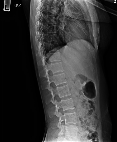
Free Images: "bestof:L1 2 vertebral fracture.jpg Skiagram kompresivní fraktury obratle L1/2 v terénu osteoporózy Lateral spine X-ray showing osteoporotic wedge fractures of L1/2"
Recursive raytracing.svg
Governor's Island.svg
L1 2 vertebral fracture.jpg
Lumbar MRI t2-tse-rst-sagittal 10.jpg
Surface projections of the organs of the trunk.png
MESI protocol activity diagram.png
Grb_profiles.gif
Cervical Xray Lateral View.jpg
Cervical Xray Lower Lateral View.jpg
Dystrophaeus_holotype.jpg
Planet Mechanics wood gasifier.png
Cervical Xray Extension.jpg
Cervical Xray Lower AP View.jpg
Cervical Xray AP View.jpg
AmiaCalvaHeadLankester.jpg
Terms of Use   Search of the Day