MAKE A MEME
View Large Image
| View Original: | Lumbar MRI t2-tse-rst-sagittal 10.jpg (799x937) | |||
| Download: | Original | Medium | Small | Thumb |
| Courtesy of: | commons.wikimedia.org | More Like This | ||
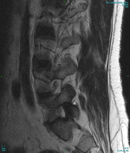
| Keywords: Lumbar MRI t2-tse-rst-sagittal 10.jpg MRI of lumbar spine T2-weighted Part of series of 12 Special note the region of hyper-intensity white oval at L5-S1 facet joint according to my spine surgeon is a lesion due to degenerative changes in the facet joint capsule Not noted on the below report but mentioned in previous studies is significant degenerative changes to the facet joints arthrosis at the L5-S1 level and also L4-L5 level to a lesser degree Radiologist's report PROCEDURE MRI of the lumbar spine without and with IV contrast CLINICAL HISTORY Lumbar pain COMPARISON 03/12/2013 TECHNIQUE Multiplanar multisequence MR imaging of the lumbar spine was performed before and after uneventful IV contrast administration 10 mm Magnevist FINDINGS The lumbar vertebral bodies are in satisfactory alignment without fracture or subluxation The vertebral body heights are maintained No abnormal bone marrow signal is identified The distal spinal cord and conus medullaris are unremarkable The paravertebral soft tissues are normal Post- contrast imaging shows no abnormal enhancement The L1-2 through L3-4 levels are unremarkable The L4-5 and L5-S1 level show stable mild degenerative changes including disc desiccation and osteophyte formation Mild disc bulges at both levels are stable both levels show mild left lateral recess narrowing with encroachment upon the transiting left L5 and S1 nerve roots respectively The minimal annular tear at L4-5 is stable as well IMPRESSION Stable mild degenerative changes and mild disc bulges at L4-5 and L5-S1 with stable minimal annular tear at L4-5 Stable mild left lateral recess narrowing at both levels with encroachment upon the transiting left L5 and S1 nerve roots respectively 2014-07-24 own Stillwaterising cc-zero Uploaded with UploadWizard Magnetic resonance imaging of the lumbar spine MRI of spinal disc herniation Facet joint disorders | ||||