Click Here for More Images from iStock- 15% off with coupon 15FREEIMAGES 
Leiden leaf c.s. under microscope
plant Zea Stem C.S. under light mircoscope with white background
Cotton plant root, under the light microscope. Longitudinal section through the root of Gossypium hirsutum, upland or also Mexican cotton. Light micrograph at 20X magnification. Isolated, over white.
Captured by a scientific microscope and Canon 5D Mark IV
forming of underskin cellulite illustration
Photomicrograph of filamentous blue-green algae, Oscillatoria species. Each band along the stand is one cell. This filament is very short probably having recently parted from a longer one. Filaments are typically many times longer. This algae is mobile, it moves and twists through the water. San Francisco Bay, California, USA. Live specimen. Wet mount, 40X objective, transmitted brightfield illumination. Note - motion blur of live specimen, very shallow depth of field, chromatic aberration and uneven focus are inherent in light microscopy.
The study of plant tissues under the microscope in the laboratory.
corn stem micrograph with dye
Epidermis. Electron microscope micrograph showing a keratinocyte of spinous layer. The epithelial cell has a polygonal shape, central nucleus with nucleolus, cytoplasm full of keratin filament bundles, and numerous dark desmosomes crossing the intercellular spaces.
Mesenchymal dysplasia. Site: Placenta
Dicot plants stem show plant vascular tissue under light microscope view for botany education.
plant rape flower cross section under light mircoscope with white background
Lime trunk
skin science 3d
Cross-section Dicot, Monocot and Root of Plant Stem under the microscope for classroom education.
Texture Pattern Design on Banknote
Microscope photo of a sambucus stem with parenchyma cells.
Fatty liver disease (steatosis) is a common condition caused by having too much fat build up in your liver. A healthy liver contains a small amount of fat. It becomes a problem when fat reaches 5% to 10% of your liver's weight.
PAPS Smear study of a young women under microscopy showing Atrophic changes in uterus which is unusual at young age.
Micro world
A 3D medical illustration of the sacrum bone isolated on a white background, showcasing its shape, structure, and anatomical features. Back view
A microscope slide showing a stained section of a human bowel, or small intestine.  It was taken through microscope lens that has a central focus through a thin layer of glass.
aquatic plant (Hornwort plant - Ceratophyllum demersum) under the microscope showing chloroplasts, cell walls and hairs - optical microscope x200 magnification
Division of Human Cancer Cells
Healthy human teeth with normal occlusion, side view. Medically accurate tooth 3D illustration
Carcinoid tumors are a type of slow-growing cancer that can arise in several places throughout your body. Carcinoid tumors, which are one subset of tumors called neuroendocrine tumors, usually begin in the digestive tract (stomach, appendix, small intestine, colon, rectum) or in the lungs. Site: Ovary
Cross section leaf of plant under the light microscope view for botany education.
Oviduct T.S. under light microscope with white background
Cross-section leaf Plant of under the microscope for classroom education.
Intricate bubble texture macro in bright colors for abstract background. Liquid bubbles lit from below with colors injected into the liquid as a design or art abstract.
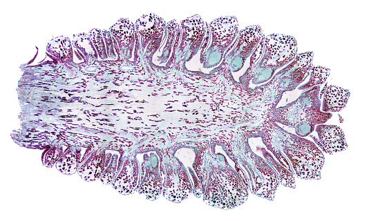
Free Images: "bestof:Gynoecium morphology placentation axile longitudinal section.png es Morfología del gineceo placentación axilar sección longitudinal Según Simpson 2005 Plant"
Plant morphology - inflorescence asteraceae compositae longitudinal section.svg
Flower morphology - longitudinal section chirimoya Annona.svg
Stem morphology type scape.svg
Flower morphology - Flower longitudinal section bilabial Bignoniaceae Tecoma stans.svg
Stem morphology type areole.svg
Stem morphology type thorn.svg
Stem morphology type culm.svg
Stem morphology type tiller.svg
Stem morphology type caudiciform stems.svg
Stem morphology type fascicle.svg
Plant morphology - inflorescence asteraceae compositae.svg
Fruit morphology - samara - flower longitudinal section wings on developing fruit Acer platanoides.svg
Gynoecium morphology style geniculate.svg
Stem morphology type tiller (modified).svg
Stem morphology type culm (modified).svg
Fruit morphology fruit type - achenecetum Fragaria.svg
Fruit morphology fruit type - drupecetum Rubus.svg
Fruit morphology fruit type - sorosis Ananas comosus.svg
Fruit morphology fruit type - achene (from multiple fruit of achenes Platanus).svg
Fruit morphology - Type bur with achenes and involucral bracts Xanthium.svg
Prunus longitudinal half cut flower ovary style stamens hypanthium.svg
Flower morphology perianth type spurred - parts calyx spur.svg
Flower morphology - longitudinal section showing achene formation and hypanthium in Rosa-01.svg
Sección_longitudinal_de_la_Iglesia_de_Torrero.jpg
Respiratory horns on the egg of Nepa Hemiptera longitudinal section after Hinton 1961.svg
Bristle morphology - plumose.svg
Bristle morphology - glochidiate.svg
Hegi_placentation.svg
Glosario del APWeb - gineceo y pistilo.svg
Flower morphology perianth type campanulate.svg
Flower morphology perianth type urceolate.svg
Flower morphology perianth type rotate.svg
Flower morphology perianth type cruciate.svg
Flower morphology perianth type infundibular.svg
Flower morphology perianth type salverform.svg
Bristle morphology - uncinate or hooked.svg
Flower morphology perianth type personate - parts constriction.svg
Flower morphology perianth type tubular part tube.svg
Bristle morphology - barbed or barbellate.svg
Flower morphology perianth type unguiculate.svg
Flower morphology perianth type saccate - parts sac or pouch.svg
Flower morphology perianth type carinate - parts keel.svg
Leaf morphology - venation Hickey 1973 - parallelodromous.svg
Leaf morphology - venation Hickey 1973 - campylodromous.svg
Leaf morphology - venation Hickey 1973 - flabellate.svg
Leaf morphology - venation Hickey 1973 - hyphodromous.svg
Leaf morphology - venation Hickey 1973 - palinactinodromous.svg
Flower morphology perianth type spurred - parts corolla spur.svg
Leaf morphology - venation Hickey 1973 - craspedodromous semi-craspedodromous.svg
Leaf morphology - venation Hickey 1973 - craspedodromous simple.svg
Leaf morphology - venation Hickey 1973 - acrodromous imperfect suprabasal.svg
Leaf morphology - venation Hickey 1973 - actinodromous imperfect marginal.svg
Leaf morphology - venation Hickey 1973 - actinodromous imperfect reticulate.svg
Leaf morphology - venation Hickey 1973 - camptodromous cladodromous.svg
Leaf morphology - venation Hickey 1973 - acrodromous perfect basal.svg
Flower morphology perianth type cucullate (hooded) - parts hood (upper lip) and lower lip.svg
Flower morphology perianth type bilabiate - parts tube throat upper and lower lip.svg
Leaf morphology - venation Hickey 1973 - camptodromous brochidodromous.svg
Leaf morphology - venation Hickey 1973 - camptodromous reticulodromous.svg
Leaf morphology - venation Hickey 1973 - acrodromous imperfect basal.svg
Leaf morphology - venation Hickey 1973 - acrodromous perfect suprabasal.svg
Leaf morphology - venation Hickey 1973 - camptodromous eucamptodromous.svg
Gray769-es.svg
Fragaria flower longitudinal section.svg
Ovule morphology anatropous.svg
Ovule morphology orthotropous.svg
Ovule morphology campilotropous.svg
Flower morphology - flower cut-away Ericaceae Arbutus.svg
Flower morphology - Flower lateral view some pieces removed - gynobasic style position epipetalous stamen Boraginaceae.svg
Juan_van_der_Hamen_y_León_-_Francisco_de_Quevedo_y_Villages.jpg
Leaf morphology ocrea.svg
Stamen morphology fusion polyadelphous.svg
portugues-sexo_alemão-sex_ingles-sex_africano-seks_russo-секс_albanes-sex_chines-性別_bulgaro-секс_croata-spol_gales-rhyw_coreano-섹스_frances-sexe_japones-セックス_filandes-sukupuolesta_arabe-الجنس_prontos_ai_tens_em_15_linguas_a_p.jpg
Style and stigma shape in color-05 - Spherical, capitate (estigma esférico, o capitado, o en cabezuela).svg
Anticlea elegans half longitudinal cut flower ovary ovules stamens style glands.svg
Densidades de población en España (2005).svg
Variación de la población española entre 1981 y 2005.svg
Quevedo.jpg
Francisco de Quevedo.jpg
Retrato_de_Francisco_de_Quevedo.jpg
Retrato_de_Francisco_de_Quevedo,_attributed_to_John_Vanderham.jpg
Quevedo_(copia_de_Velázquez).jpg
Oceanic ecosystemes.svg
Araya,_Agustin_(orig_de_Blanes)_-_Los_ultimos_momentos_de_JM_Carrera.jpg
Escudo de España.svg
Bandera de España (construccion).svg
Design_Analysis_of_the_P-38_Lightning.png
Design_Analysis_of_the_Boeing_B-17G_Flying_Fortress.png
Design_Analysis_of_the_Lockheed_PV-1_Ventura.png
Flag of Spain and Mexico.png
Gynoecium morphology placentation axile longitudinal section.png
Gynoecium morphology placentation apical-axile longitudinal section.png
Gynoecium morphology placentation axile-1 cross section.png
Gynoecium morphology placentation axile-2 cross section.png
Gynoecium morphology placentation parietal-axile cross section.png
Gynoecium morphology placentation basal longitudinal section.png
Gynoecium morphology placentation marginal longitudinal section.png
Gynoecium morphology placentation apical-pendulous longitudinal section.png
Terms of Use   Search of the Day