Click Here for More Images from iStock- 15% off with coupon 15FREEIMAGES 
Plant cells under microscope
plant Tilia Stem C.S. under light mircoscope with white background
The study of plant tissues under the microscope in the laboratory.
Embryonic Stem Cells (ES cells) under microscope.
Green illuminate glitter bokeh for background for concept and more idea for creative
Leiden leaf c.s. under microscope
Moss leaf under the microscope, green cell background
Beautiful cellular pattern, vegetal tissue with special dye.
Aerial view of loyster farm by Ardara, County Donegal - Ireland.
Particles, medical concept
Pine mature wood cross section. Light microscope slide with microsection of an evergreen conifer in the genus Pinus. Plant anatomy. Biology. Photo.
Viral Infection Monkeypox Virus - 3d rendered image. Abstract biomedical illustration. \nAntibody, Antigen, Vaccine technology concept.
Microbial Biofuel energy cell in macroscopic view. Concept 3D illustration mockup. Solar-to-fuel algae Ethanol power and engineered cyanobacterium, algal bacteria as future zero waste renewable energy
Abstract red and green spotted virus pattern background
Cross-section Dicot, Monocot and Root of Plant Stem under the microscope for classroom education.
Close up of cristal beads
Light photomicrograph of pine tree wood seen through a microscope
Phospholipid bilayer membrane structures that make up living cells and their contents. This is an image of membrane proteins in the membrane.\nThis image, which looks like a plastic model, evokes biology and biochemistry. It can be used as a cover for magazines, textbooks and presentation materials.\nThis image was not created by AI.
Microscopic view of a colony of Aspergillus fungi, which causes the lung infection aspergillosis, aspergilloma of the brain and lungs. 3D illustration
corn stem micrograph with dye
Microalgae under microscopic view, green algae, cyanobacteria, phytoplankton, diatom, algae mix collage background
Host cells with spores (mold) are inside wood under the microscope for education.
Abstract blue cell pattern, spots, cool, virus, background effect, texture
False colour transmission electron microscope (TEM) micrograph of a mast cell (mastocyte) with the cytoplasm (green) full of very large histamine/heparin granules (dark blue). Nucleus is brown.
Endosomes - 3d rendered image of Endosomes are membrane-bound organelles. They are parts of endocytic membrane transport pathway.  Endomembrane system in cells. Bilayer Lipid Membrane. Abstract medical illustration. Microscopic view. Microbiologic research concept.
Blue colored stem cells of a lentil plant under the microscope.
Different germs in the human intestines called microbiome,Bacteria Lactobacillus in human intestine,Beneficial healthy intestinal bacterium microflora,Gut bacteria
Cross section - Phloem is a type of tissue in vascular plants that transports water and some nutrients. Scientific research. Plant tissue Structure.
Line Pattern Design on Banknote
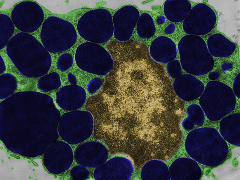
Free Images: "bestof:Colorized scanning electron micrograph of filamentous Ebola virus particles (green) attached to and budding from a chronically infected VERO E6 cell (blue)"
Ebola_Virus_Particles.jpg
Ebola_virus_particles_found_both_as_extracellular_particles_and_budding_particles_from_chronically-infected_African_green_monkey_kidney_cells_..jpg
hela-cells-bacteria-germs-virus-544318.jpg
tin-spheres-colorized-1444073.jpg
Hepatitis B virus v2.svg
Malaria-infected_Red_Blood_Cell.jpg
H1N1_virus_particles.jpg
Cube of Germanium (5828204117).jpg
H1N1_virus_particles_II.jpg
Multidrug-resistant Klebsiella pneumoniaeand neutrophil.bmp.jpg
Red_blood_cell_infected_with_malaria_parasites-NIAID_RML.jpg
Cygnet_River_square_(EID_2012_Fig1a).jpg
Hhv-6 (1).jpg
Bone Cells (5828753702).jpg
Giardia lamblia.bmp.jpg
HIV_H9_T-cell_II.jpg
White_blood_cell_with_MRSA.jpg
Electron_Micrograph_of_Flea_derivate.jpg
Diseases of Swine 23-3.png
Staph_aureus_outside_a_white_blood_cell.jpg
Scanning_Electron_Micrograph_of_a_Flea.jpg
Giardia_1.jpg
SARS_CoV.jpg
Tin Spheres (5828754970).jpg
HeLa-IV.jpg
TEM_phil_CDC_11635_lores.jpg
HeLa-V.jpg
Neutrophil_MRSA_I.jpg
Streptococcus_pyogenese_Bacteria.jpg
Chikungunya_virus_particles-PHIL-17369.jpg
Human_neutrophil_ingesting_MRSA.jpg
Neutrophil_MRSA_II.jpg
Chlamydomonas6-1.jpg
Velvet_ant_head_anterior_--_SEM_photo.jpg
B00528-Swine-flu.png
Surface_of_HIV_infected_microphage.jpg
Human Lung Cells (6680207077).jpg
New Quantum Dot Transistor Counts Individual Photons (5941062028).jpg
Macrophages in the brain before and after in vitro infecti.jpg
Embryonic_smooth_muscle_cell.jpg
Chemical Analysis;Maximum Pixel Software (5884305705).jpg
Cerebellum PurkinjeCells.jpg
LDLR pathway.png
Avoid_buying_these_tables_for_home_or_business_(1).jpg
Avoid_buying_these_tables_for_home_or_business_1.jpg
Hhv-6.jpg
Red_White_Blood_cells.png
Head of Chrysomelidae derivat.jpg
Terms of Use   Search of the Day