Click Here for More Images from iStock- 15% off with coupon 15FREEIMAGES 
Blue colored stem cells of a lentil plant under the microscope.
Natural agate surfaces, suitable for backgrounds and wallpapers.
Epidermis. Electron microscope micrograph showing a keratinocyte of spinous layer. The epithelial cell has a polygonal shape, central nucleus with nucleolus, cytoplasm full of keratin filament bundles, and numerous dark desmosomes crossing the intercellular spaces.
Blastomycosis is a fungal infection caused by inhaling Blastomyces dermatitidis spores. If it involves only the lungs, it is called pulmonary blastomycosis. Only about half of people with the disease have symptoms, which can include fever, cough, night sweats, muscle pains, weight loss, chest pain, and feeling tired.
Tissue of Small intestine (Duodenum), Large intestine Human and Stomach Human under the microscope in Lab.
Photomicrograph of two rotifers. Rapidly rotating cilia at top of heads, internal organs visible. Live specimen. Wet mount, 10X objective, transmitted brightfield illumination.
centropyxis aculeata (amoeboid organism) under the microscope - optical microscope x400 magnification
corn stem micrograph with dye
Microscopic photo of a thin section of calcareous tufa of Holocene age.
Colorful seamless pattern with Crystal stones.
Cross sections of the plant root under the microscope view for education
Particles, medical concept
the tail of Aelurostrongylus abstrusus, cat lungworm under the microscope
Equisetum is a \
Transmission electron micrograph (TEM) showing a desmosome (macula adherens) with prominent dense plaques where keratin intermediate filaments were attached.
Diffuse large B-cell lymphoma, or DLBCL, is a cancer that starts in white blood cells called lymphocytes.
Microscope photo of a sambucus stem with parenchyma cells.
Pine Root C.S. under light mircoscope with white background
The study of plant tissues under the microscope in the laboratory.
Cheyletiella blakei Mite - Small Cat pest view under a microscope
Kidney cancer: Clear cell carcinoma. fuhrman nuclear grade-II, Renal growth shows a malignant tumor, renal cell carcinoma in between renal tubules.
Photomicrograph of flatworm,
Enlarge microscopic view of larvae stage of crustacean
Neuroendocrine carcinoma well differentiated (carcinoid) rectum with immunostain synaptophysin
Notoedres cati under the microscope. Notoedric mange, also referred to as Feline scabies, is a highly contagious skin infestation caused by an ectoparasitic and skin burrowing mite Notoedres cati.
Curly marble stone with fashion veins.
microscopy micrograph plant tissue, stem of pumpkin, magnification 200X
Liver Fluke Section under light microscope with white background
Stratified flat epithelium under microscope
A model of the phospholipid bilayer structure that makes up living cells and their contents. Its raw, beautiful appearance evokes images of biology and biochemistry. It can be used in magazines and textbooks and on the covers of presentations.\nThis image was not created by AI.
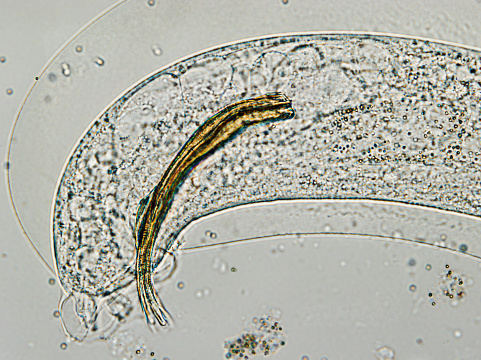
Free Images: "bestof:Schistosoma mansoni egg 4841 lores.jpg This micrograph depicts an egg from the parasite Schistosoma mansoni and reveals the egg �s characteristic lateral spine"
Mesoplia_aff_rufipes,_F,_Face,_Puerto_Rico,_Boqueron_2014-09-26-09.36.28_ZS_PMax.jpg
Mesoplia_aff_rufipes,_M,_back,_Puerto_Rico,_Yauco_2014-09-26-10.00.23_ZS_PMax.jpg
Mesoplia_aff_rufipes,_F,_Back,_Puerto_Rico,_Boqueron_2014-09-26-09.27.04_ZS_PMax.jpg
Mesoplia_aff_rufipes,_M,_face,_Puerto_Rico,_Yauco_2014-09-25-10.33.22_ZS_PMax.jpg
Mesoplia_aff_rufipes,_M,_side,_Puerto_Rico,_Yauco_2014-09-26-10.22.25_ZS_PMax.jpg
Mesoplia_aff_rufipes,_F,_Side,_Puerto_Rico,_Boqueron_2014-09-26-09.45.54_ZS_PMax.jpg
Physocephala_tibialis,_back,_scarpulla_,_virginia_2014-07-09-18.06.jpg
Physocephala_tibialis,_face,_scarpulla,_virginia_2014-07-09-18.19.jpg
Physocephala_tibialis,_side,_scarpulla,_vriginia_2014-07-09-18.25.jpg
Pasites_maculatus,_f,_pakistan,_back_2014-11-02-01.39.25_ZS_PMax.jpg
Pasites_maculatus,_f,_pakistan,_side_2014-11-02-01.50.40_ZS_PMax.jpg
Pasites_maculatus,_f,_pakistan,_face_2014-11-02-01.46.02_ZS_PMax.jpg
Pieter_Brueghel_the_Elder_-_The_Dutch_Proverbs_-_Google_Art_Project.jpg
Schistosoma life cycle.svg
Clonorchis sinensis LifeCycle-french.svg
Schistosoma_mansoni_egg_4841_lores.jpg
Schistosoma_haematobium_egg_4842_lores.jpg
Giardia_1.jpg
S_mansoni_eggF.jpg
Schistosoma mansoni2.jpg
Trichuris_trichiura_egg.tif
S_interculatum_eggS.jpg
S_mekongi_eggR.jpg
Eggs_of_Trichuris_trichiura_and_Trichuris_vulpis_06G0018_jpg_lores.jpg
FMIB_47897_Fall_Web_Worm_and_Spiny_Elm_Caterpillar-_FAll_Web_Worm_(Hyphantria_cunea_Drury)-_1_Cluster_of_eggs;_2_Dorsal_views_of_full_and.jpeg
S_mansoni_eggT.jpg
Schistosoma 20041-300.jpg
Egg_of_Fasciola_hepatica_08G0041_lores.jpg
Embryonated_chicken_egg-10759_lores.jpg
Psychosynthesis-egg-diagram.png
Phyllium giganteum - egg from lateral.jpg
Phaenopharos khaoyaiensis Egg.jpg
FMIB_39995_Anchovia_mitchilli-_Egg_showing_advanced_stage_in_development_of_embryonic_shield,lateral_view,_gr,_germ_ring.jpeg
FMIB_40000_Bairdiella_chrysura-_Egg_with_blastoderm_of_may_cells,_late_cleavage_stage,_lateral_view;_pb,_periblast.jpeg
Taenia_solium_tapeworm_proglottids_5261_lores.jpg
House_of_Fabergé_-_Gatchina_Palace_Egg_-_Walters_44500_-_Closed.jpg
House_of_Fabergé_-_Gatchina_Palace_Egg_-_Walters_44500_-_Open_View_B.jpg
Dipyl_can_egg1.JPG
Terminal_chlamydospores_of_the_fungus_Microsporum_audouinii_PHIL_3054_lores.jpg
Gravid_proglottid_from_the_cestode_Taenia_saginata_5259_lores.jpg
Zp3 structure 3d4g.png
2011-LHayes-proof-rev.jpg
Cargese_Greek_Church_Three_Hierarchs.jpg
Cargese_Greek_Church_John_the_Baptist.jpg
Illustrated_Life_Cycle_of_Echinostoma.gif
FMIB_47740_Apeltes_quadracus_(Four-spined_Stickleback).jpeg
Egyptian - Head of a Priest - Walters 2263.jpg
Giardia lamblia.bmp.jpg
Scabies_LifeCycle(French_version).GIF
Cassel_Painter_-_Kalpis_with_Toilette_Scene_with_Two_Women_and_Eros_-_Walters_4878_-_Bottom.jpg
Cassel_Painter_-_Kalpis_with_Toilette_Scene_with_Two_Women_and_Eros_-_Walters_4878_-_Side_A.jpg
Cassel_Painter_-_Kalpis_with_Toilette_Scene_with_Two_Women_and_Eros_-_Walters_4878_-_Side_A_Detail.jpg
Aoki_Harutsura_-_Kozuka_with_a_Horse_and_Show_Monkey_-_Walters_51634_-_Back.jpg
Aoki_Harutsura_-_Kozuka_with_a_Horse_and_Show_Monkey_-_Walters_51634.jpg
Aoki_Harutsura_-_Kozuka_with_a_Horse_and_Show_Monkey_-_Walters_51634_-_Mark_A.jpg
Bowdoin_Painter_-_Shoulder_Lekythos_Depicting_Nike_(?)_Pouring_from_a_Patera_at_an_Altar_-_Walters_48257_-_Right_Side.jpg
Bowdoin_Painter_-_Shoulder_Lekythos_Depicting_Nike_(?)_Pouring_from_a_Patera_at_an_Altar_-_Walters_48257_-_Detail.jpg
Bowdoin_Painter_-_Shoulder_Lekythos_Depicting_Nike_(?)_Pouring_from_a_Patera_at_an_Altar_-_Walters_48257_-_Side_A.jpg
Parapet and Cornice at 102 Marine Parade, Brighton.JPG
Epimedes_Painter_-_Amphora_with_a_Youth_and_a_Bearded_Man_and_Victory_-_Walters_4853_-_Side_A.jpg
Epimedes_Painter_-_Amphora_with_a_Youth_and_a_Bearded_Man_and_Victory_-_Walters_4853_-_Side_A_Detail.jpg
Epimedes_Painter_-_Amphora_with_a_Youth_and_a_Bearded_Man_and_Victory_-_Walters_4853_-_Side_B.jpg
Epimedes_Painter_-_Amphora_with_a_Youth_and_a_Bearded_Man_and_Victory_-_Walters_4853_-_Side_B_Detail.jpg
Patanazzi_family_-_Ewer_Basin_with_Leda_and_the_Swan_-_Walters_481510.jpg
FMIB_47898_Simulium_Society-_1_Two_imagos_of_hydropsyche;_2_Imago_of_hydorpsyche;_3_Larva_of_hydropsyche;_4_Pupal_case_of_hydropsyche;_5.jpeg
F._W._Bolgiano,_seedsmen_have_won_confidence_for_ten_years_(15751236504).jpg
Wholesale_price_list_of_requisites_for_florists_and_market_gardeners_(16569158317).jpg
Wholesale_price_list_of_requisites_for_florists_and_market_gardeners_(16775279591).jpg
Dreer's_wholesale_price_list_(16166193413).jpg
Wholesale_price_list_of_requisites_for_florists_and_market_gardeners_(16776427505).jpg
Garden_and_farm_seeds_(15752666413).jpg
Bulbs_and_plants_(16200770938).jpg
Terms of Use   Search of the Day