Click Here for More Images from iStock- 15% off with coupon 15FREEIMAGES 
Proteasomes are molecular machines for breaking down proteins called proteolysis, only target proteins that have been marked for destruction called ubiquitin to the target protein, 3d rendering
Global communication and collaboration intricately involving electronic ropes and keys through social networking of smart phones equipped with AI.
3D cartoon and Gaussian surface models, PDB 6vg8, white background
The three-dimensional structure of proteins is represented in a variety of conformations. This image is based on the ribbon structure of a protein. It is suitable for biology notebooks and book covers.\nThis image has not been created by AI.
After dopamine binding, the dopamine receptor changes his shape and binds the inactive G protein.The G protein expels its GDP molecule replacing it by GTP. and falls into two pieces. The Galpha subunit binds to and activates the enzyme adenylyl cyclase, which produces then cyclic AMP spreading a signal through the cell.
A ball-and-stick plus ribbons molecular model of the hormone Orexin, also known as Hypocretin.  Orexin is involved in the regulation of the sleep/wake cycle and it stimulates apetite, wakefulness and energy expenditure. It has been linked with Alzheimer's disease.  Isolated on white.
3d rendered Gastric inhibitory polypeptide (GIP, glucose-dependent insulinotropic peptide) endocrine protein hormone, 3D illustration
3d rendering of transparent molecule shape on green color background.
DNA mutation, a mutation is a change in a DNA sequence, mutations can result from DNA copying mistakes made during cell division.
Cells in the body examined by endoscope
Resistin also known as adipose tissue-specific secretory factor (ADSF) or C/EBP-epsilon-regulated myeloid-specific secreted cysteine-rich protein (XCP1) is a cysteine-rich peptide hormone derived from adipose tissue that in humans is encoded by the RETN gene. 3D cartoon and Gaussian surface models, chain id color scheme, based on PDB 1rfx, white background
Phospholipid bilayer membrane structures that make up living cells and their contents. This is an image of membrane proteins in the membrane.\nThis image, which looks like a plastic model, evokes biology and biochemistry. It can be used as a cover for magazines, textbooks and presentation materials.\nThis image was not created by AI.
Amitriptyline is used as an antidepressant and antimigraine drug. It blocks sodium channels on neuronal membrans. Sodium channels generate and propagate action potentials in neurons and muscle. Source: PDB entry 6AGF
Covid-19 Coronavirus Variants Omicron Delta Gamma Beta Alpha Pandemic Virus Disease Mutation
Cell Membrane and Molecules, 3d rendering. Computer digital drawing.
Abstract spheres molecular structure neural network.  Blockchain and artificial intelligence concept. 3d render.
Multicolored antibodies or immunoglobulin protein structures - 3d illustration
3d Rendering Molecular Structure,Ozone,Hydrogen and oxygen
3D cartoon and Gaussian surface models, PDB 2gjx, white background
Inside protein molecule
Bacterial multidrug efflux pumps (E-CmeB) expel antibiotics  from inside the bacterium (bottom): ciprofloxacin (yellow), chloramphenicol (violet),  ampicillin (red).
A ball and stick model of a strand of a molecule of polyvinyl chloride, more commonly known as PVC. This vinyl polymer is one of the commonest plastics in use.
Perfluoroisobutene fluorocarbon molecule.  3D rendering. Atoms are represented as spheres with conventional color coding: carbon (grey), fluorine (lig
Digital background depicting innovative technologies in (AI) artificial systems, neural interfaces and internet machine learning technologies
proteins as analytes conjugate on antibody molecule or receptor flow through metal film nanoholes of opto-fluidic biosensor 3d rendering
Endosomes - 3d rendered image of Endosomes are membrane-bound organelles. They are parts of endocytic membrane transport pathway.  Endomembrane system in cells. Bilayer Lipid Membrane. Abstract medical illustration. Microscopic view. Microbiologic research concept.
3D cartoon and Gaussian surface models, chain id color scheme, PDB 3omv, white background
Neural Network Nodes Deep Learning Artificial Intelligence Machine Learning Model
The calcitonin gene related peptide (yellow) binds to its receptor (blue) on neurons and smooth muscle cells of cerebral blood vessels, activating a signal cascade through G-proteins (dark blue) in this cells that  leads to a dilatation of the blood vessels (vasodilatation).
3d rendering of Easter eggs background
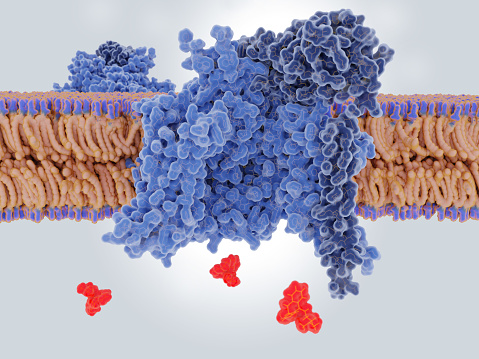
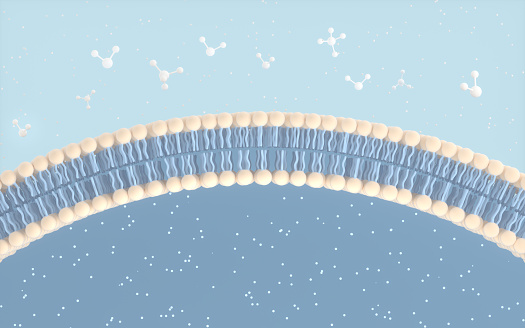
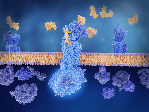
Free Images: "bestof:Scheme facilitated diffusion in cell membrane-zh.svg Facilitated diffusion involves the use of a protein to facilitate the movement of molecules across the"
Scheme facilitated diffusion in cell membrane-zh.svg
Scheme facilitated diffusion in cell membrane-zh (zh-tw).svg
Scheme facilitated diffusion in cell membrane-en.svg
Scheme facilitated diffusion in cell membrane-eo.svg
Scheme facilitated diffusion in cell membrane-ca.svg
Scheme simple diffusion in cell membrane-fr.svg
Scheme facilitated diffusion in cell membrane-fr.svg
Scheme simple diffusion in cell membrane-zh (zh-cn).svg
Scheme simple diffusion in cell membrane-zh (zh-tw).svg
Scheme simple diffusion in cell membrane-en.svg
Scheme facilitated diffusion in cell membrane-es.svg
Scheme simple diffusion in cell membrane-es.svg
Scheme simple diffusion in cell membrane-pl.svg
Scheme simple diffusion in cell membrane-de.svg
Scheme simple diffusion in cell membrane-ca.svg
Scheme simple diffusion in cell membrane-de 02.svg
Scheme secundary active transport-heb.svg
Scheme secundary active transport-en.svg
Scheme secundary active transport-pl.svg
Scheme sodium-potassium pump-ja.svg
Scheme sodium-potassium pump-it.svg
Scheme sodium-potassium pump-en-2.svg
Scheme sodium-potassium pump-zh.svg
Scheme sodium-potassium pump-zh (zh-tw).svg
Scheme sodium-potassium pump-en.svg
Scheme sodium-potassium pump-pl.svg
Scheme sodium-potassium pump-bs.svg
Scheme sodium-potassium pump-he.svg
Scheme sodium-potassium pump-el.svg
Scheme sodium-potassium pump-de.svg
Scheme sodium-potassium pump-ca.svg
Scheme sodium-potassium pump-cs.svg
Design_Analysis_of_the_B-24J_Liberator.png
portugues-sexo_alemão-sex_ingles-sex_africano-seks_russo-ÑекÑ_albanes-sex_chines-性別_bulgaro-ÑекÑ_croata-spol_gales-rhyw_coreano-섹스_frances-sexe_japones-セックス_filandes-sukupuolesta_arabe-الجنس_prontos_ai_tens_em_15_linguas_a_p.jpg
Scheme facilitated diffusion in cell membrane-de.png
Scheme simple diffusion in cell membrane-he.png
Scheme simple diffusion in cell membrane-de.png
Porters.PNG
DiffusionMicroMacro.gif
NIST physicist Michael Boss (16775001192).jpg
3D Test Method for Biomaterials (5885545588).jpg
Junctional complex and pinocytotic vesicles - embryonic brain-TEM.jpg
Unctional complex and pinocytotic vesicles - embryonic brain - TEM.jpg
Gag Molecules (5888007689).jpg
Nanopore-based single molecule mass spectrometry (5884864158).jpg
DNA_Repair.jpg
V135 sherbrooke arms.jpg
Terms of Use   Search of the Day