Click Here for More Images from iStock- 15% off with coupon 15FREEIMAGES 
An ultra sound of a baby.
Collection of  MRI heart or Cardiac MRI ( magnetic resonance imaging ) of heart  showing heart beating for detecting heart disease.
Doctor looking at his patients X-ray image on digital tablet
Ultrasound image of myoma or uterine fibroids. Medical research
Doctor holds ultrasonic probe in hand preparing device for examination. Ultrasound of internal organs concept
Medical imaging of various stages of embryo and fetal development during pregnancy. Progression includes:
Ultrasound examination of the abdominal cavity.
Four, side view, X-ray pictures of a woman's two breasts. On the right, lateral breast there are two intramammary lymph nodes with fatty hilum measuring 0.5 and 0.6 cm respectively. No suspicious breast mass or auxiliary lymphadenopathy. Moderately dense breasts with 50 to 75 % glandular tissue.  Bi-RADS 2 : benign findings.
Cropped image of screen showing researcher looking in a microscope fertilising an egg. Intracytoplasmic sperm injection.
Ultrasound of Kidney  or KUB  for  screening  renal stone disease or Urolithiasis.
Sonography examination of cystic thyroid nodule showing the solid nodule embedded in normal thyroid tissue
Two computer monitors are seen inside a CT scan control room
Photos of the child in the 12 week of pregnancy. Ultrasound procedure. Screening in the first trimester
X-ray of female hips, pelvis and spine
CT Abdomen Cross section View. CT scan (computed tomography) of chest organs.
Close-up of a x-ray image displayed on computer on tomography room
Full size digital screening mammogram of a new breast tumor in the retroareolar region of the right breast causing nipple retraction. An ultrasound guided biopsy was performed diagnosing invasive ductal carcinoma. This is the most common type of breast cancer, accounting for nearly 80% of cases. Breast cancer treatment has come a long way in the last few decades, seeing more funding than any other human disease. The first step to a cure is catching it early with screening mammograms such as this case so that the tumor can be removed before it spreads (metastasizes) out of the breast.
MRI of the prostate gland, revealing an enlarged size, aids in diagnosing tumors, guiding treatment decisions, and monitoring prostate health.
Human Skeletal System X-Ray Images for Medical Education
Computed tomography of chest organs. CT scan. CT Abdomen Cross section View.
CAT Scan of a woman with left breast cancer showing an increase between the skin and the prosthesis Medical themes
Ultrasound image of a breast
x-ray images of urinary bladders or bladder and prostate, gland or prostatic for check Prostatic diseases or Prostatic hyperplasia.
Computational tomography image of the abdominal and pelvic regions.
Anatomy of the pelvis bones, including the ilium, ischium, sacrum, and pubis, photorealistic 3D illustration. Front view. Perfect for educational or medical purposes
X-ray of the human mouth showing dental
MRI heart or Cardiac MRI ( magnetic resonance imaging ) of heart compare RVOT and LVOT for diagnosis heart disease.
Wide view of CT scanner room
Nine images of fetus ultrasonography.
MRI scanner machine in medical examination room
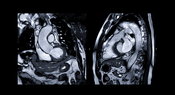
Free Images: "bestof:RenalCancer-90MBq-F-18-Fluorid-PET-CT Vergleich.jpg en Sodium fluoride PET/CT Scan of a bone metastasis of kidney cancer; the PET scan shows the metabolic"
Albrecht_Dürer's_"young_hare".jpg
Fully_Integrated_Whole-body_Simultaneous_PET/MRI_Device.jpg
PET-MIPS-anim.gif
Edward_Walsh_-_Queenstown,_Upper_Canada_on_the_Niagara_(a.k.a._Queenston,_Ontario).jpg
Symptoms of cancer metastasis.svg
Målning._På_besök_hos_konsthandlaren._Frans_Francken_II_-_Hallwylska_museet_-_86711.tif
World oil consumption 1980 to 2007 by region.svg
Oil consumption per day by region from 1980 to 2006 solid2.svg
Oil consumption per day by region from 1980 to 2006 solid3.svg
Metastasis sites for common cancers-IT.svg
HolidaysWeedsHaveMoreFunThanMartinsHorses.jpg
RenalCancer-90MBq-F-18-Fluorid-PET-CT Vergleich.jpg
RenalCancer-250MBq-FDG-PET-CT Vergleich.jpg
RenalCancer-90MBq-F-18-Fluorid-PET-CT.jpg
RenalCancer-250MBq-F-18-FDG-PET-CT.jpg
Knochenmetastase.jpg
PET_scan-normal_brain-alzheimers_disease_brain.PNG
PET scan image.jpg
Struma maligna.jpg
HodgkinLymphomPETCT.jpg
PET_Alzheimer.jpg
PET_Normal_brain.jpg
Dr._Nora_Volkow_with_patient_in_PET_scan.jpg
Pem mammography.jpg
PET physiologic muscle activity.jpg
Blasentumor.jpg
Constance_Crawley_-_1908.jpg
Cinq_Semaines_en_ballon_021.png
Cinq_Semaines_en_ballon_033.png
3D rendered CT of hip bone metastases.jpg
PET scans of brain tumor using Sigma-2 Ligands.png
Petofi 1848 marcius 15.jpg
Bod_-_Historia_Hungarorum_ecclesiastica_-_1888_-_Universiteitsbibliotheek_VU_XU.05390.JPG
Jablonczay_Pethes_János_jegyző_zsiványnyelv.jpg
Metastasis sites for common cancers-IT.png
Niersteen_en_mes_1740_Jan_Lethaus.JPG
Maudslays'_Map_of_Ixkun_1899.jpg
Maudslay's_1887_Map_of_Ixkun,_enhanced.png
The_dogs_of_war,_Saving_lives_but_paying_the_price_120404-N-UR169-007.jpg
Reich_Péter_Cornel_-_Dunapart,_1937._október_10_(1).tif
PET_scan_of_human_brain.png
pet_scan.png
scan_2.jpg
scan_10.jpg
Bone scan different resolutions.jpg
Gastritis PET.jpg
VuePointHD.jpg
Histograms of Bone Scan and Radiograph.jpg
Umbrella Cockatoo.jpeg
RectalCancerStagingInPETCT+arrow.jpg
BoneScanOfAHand32x32x8Bits.png
BoneScanOfAHand256x256x8Bits.png
BoneScanOfAHand256x256x2Bits.png
RitaB.jpg
Pet. van Mastricht.jpg
PET-schema.png
PetTheory2013.jpg
Histograms of pixel values in a bone scan and radiograph.gif
Histograms of pixel values in a bone scan and radiograph.jpg
TwoHandsGreyscale.jpg
Hufeisenniere VR.jpg
dear_happiness_058.jpg
CTWristImage.png
NM16 42a.jpg
NM13 33.jpg
Pet-coke-pile-with-car-20140329-fa.jpg
CT of hip bone metastases.jpeg.jpg
NM13 34.jpg
CaCa Bleu (Blå Skåpet) "Krismäss,uj!" 2009.jpg
Point frederick.JPG
005_Diriks_Skisser.jpg
Abbots Bromley original hobby horse.jpg
Face_recognition.jpg
CaCa Bleu (Blå Skåpet) "Pardon Me for Loving & Running!" 2009.jpg
Psittacus erithacus timneh -upper body-8a.jpg
Maltoid_milk-bone.jpg
CaCa Bleu (Blå Skåpet) "Kom & lek hemma hos mig, du!" 2010.jpg
US_Navy_100216-N-8335D-113_hief_Mineman_William_Brown_shows_Royal_Cambodian_Navy_officers_a_Klein_side-scan_sonar_aboard_the_mine_counter-measures_ship_USS_Patriot_(MCM_7).jpg
CT pericardial effusion.jpg
'Exotic ultra face Persian cat' at 'Mumbai cat show-2014'.JPG
Meißen_Buschbad_um_1830_(01).jpg
August_Scherl_Verlag_1914.jpg
Arolsen_Klebeband_01_055.jpg
Arolsen_Klebeband_01_115.jpg
The_Quarrel_of_Oberon_and_Titania_(detail).jpg
USS_Blueback_SS581_page_from_decommissioning_booklet.jpg
Arolsen_Klebeband_02_359.jpg
Ljubljana 115 02 1641.jpg
Ljubljana 115 03 1641.jpg
US_Navy_050423-N-8629M-073_Lt._Steve_Ferrara_shows_distinguished_visitors_from_Indonesia_the_Computed_Axial_Tomography_(CAT)_scan_capabilities_aboard_the_Military_Sealift_Command_(MSC)_hospital_ship_USNS_Mercy_(T-AH_19).jpg
US_Navy_110427-N-2531L-135_Tori_Randall,_Ph.D._prepares_a_550-year_old_Peruvian_child_mummy_for_a_CT_scan.jpg
Refresh scan.jpg
Netleysamnathbuck.jpg
Kasberg-map-1822-greyscales.jpg
Oplontis_Fig_basket.jpg
Wilms Tumor CTScan.gif
Elisabeth roses.jpg
Hamartoma of the spleen.jpg
Swaziland Visa and stamps.jpg
Smyrne Group of Gypsy.jpg
Terms of Use   Search of the Day