MAKE A MEME
View Large Image
| View Original: | PET-schema.png (1280x938) | |||
| Download: | Original | Medium | Small | Thumb |
| Courtesy of: | commons.wikimedia.org | More Like This | ||
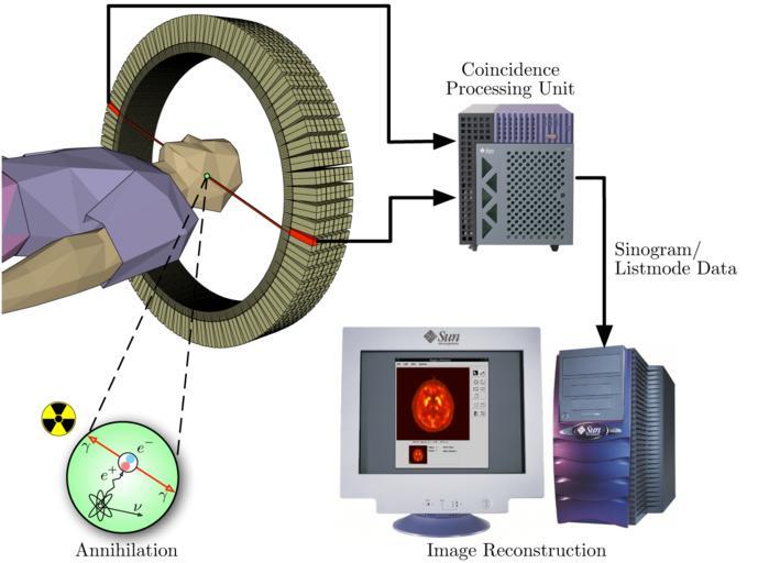
| Keywords: PET-schema.png The image illustrates the processing principles of a positron emission tomograph PET commonly used in cancer diagnostics It shows how during the annihilation process two photons are emitted in diametrically opposite directions These photons are registered by the scanner as soon as they arrive at the detector ring After the registration the data is forwarded to a processing unit which decides if two registered events are selected as a so-called coincidence event All coincidences are forwarded to the image processing unit where the final image data is produced via mathematical image reconstruction procedures own work - part of master thesis http //jens-maus de/ftp/langner_mscthesis pdf http //jens-maus de/ftp/langner_mscthesis bib 2003-04 Jens Maus http //jens-maus de/ Positron emission tomography | ||||