Click Here for More Images from iStock- 15% off with coupon 15FREEIMAGES 
Motor Neuron cells under the microscope
plant Zea Stem C.S. under light mircoscope with white background
Backgrounds of Bacteria and Mold under the microscope in Lab.
Plasmodium falciparum is the most dangerous form of malaria. Numerous parasites have infected the red blood cells of this patient. The parasites can be seen as little rings inside the red blood cells in.this thin blood smear.  Malaria is spread by mosquitoes.
Bacteria of different shapes, cocci and rod-shaped bacilli, 3D illustration in old book vintage style
Photomicrograph of filamentous blue-green algae, Oscillatoria species. Each band along the stand is one cell. This filament is very short probably having recently parted from a longer one. Filaments are typically many times longer. This algae is mobile, it moves and twists through the water. San Francisco Bay, California, USA. Live specimen. Wet mount, 40X objective, transmitted brightfield illumination. Note - motion blur of live specimen, very shallow depth of field, chromatic aberration and uneven focus are inherent in light microscopy.
Freshwater Closterium algae (unicellular charophyte green algae) - optical microscope x200 magnification
corn stem micrograph with dye
Microalgae under microscopic view, green algae, cyanobacteria, phytoplankton, diatom, algae mix collage background
Diffuse large B-cell lymphoma, or DLBCL, is a cancer that starts in white blood cells called lymphocytes.
the tail of Aelurostrongylus abstrusus, cat lungworm under the microscope
Natural agate surfaces, suitable for backgrounds and wallpapers.
Cedar pollen
Close up of small feeding tick with shiny pinhead. Nikon D7200 with Nikon 105mm macro lens & extension tube, SB 21 flash. Photo taken in Gainesville, Florida
Eggs of helminths Enterobius vermicularis containing worm larva. Threadworm which cause enterobiasis, 3D illustration
Spider and its prey isolated on white. Studio shot.
Cross section leaf of plant under the light microscope view for botany education.
Burkitt lymphoma is a form of non-Hodgkin's lymphoma in which cancer starts in immune cells called B-cells. Recognized as a fast growing human tumor, Burkitt lymphoma is associated with impaired immunity and is rapidly fatal if left untreated.
centropyxis aculeata (amoeboid organism) under the microscope - optical microscope x400 magnification
Photomicrograph of the ciliate Blepharisma americanum in various stages of development. Typical faint pink color. Each individual is only one cell. Live specimen. Wet mount, 10X objective, transmitted brightfield illumination.
Striped mosquitoes life circle with white background. Life cycle of mosquitos isolate on white background.
Squash preparation of  onion root tip cells stained to reveal chromosomes,  Bright field illumination, light microscopy.  This image was taken with the X40 objective lens.  Depth of focus is extremely low at this magnification.  Smaller apertures give greater depth of focus, but image quality deteriorates and resolution is greatly reduced.  The aperture in this image was optimised for contrast and resolution. Chromatic abberation is inevitable in light microscopy, but has been kept to a minimum in this image.
Microscopic close view of high vaginal swab Gram stain smear, 100x. diagnosis of Bacterial vaginosis (BV).
Human Morula Cell, Solid Ball of Cells Resulting from Division
newly hatched freshwater fish under a light microscope, isolated on white
Liver Fluke Section under light microscope with white background
Green algae cells background
Myxoid liposarcoma.  Myxoid/round cell liposarcoma, or MRCLS, is one of several types of liposarcoma. Liposarcoma is a rare cancer that grows in the cells that store fat in the body. MRCLS usually grows in the arms and legs. These tumors grow slowly, and they can spread to other parts of the body
Microscope photo of a sambucus stem with parenchyma cells.
Endosomes - 3d rendered image of Endosomes are membrane-bound organelles. They are parts of endocytic membrane transport pathway.  Endomembrane system in cells. Bilayer Lipid Membrane. Abstract medical illustration. Microscopic view. Microbiologic research concept.
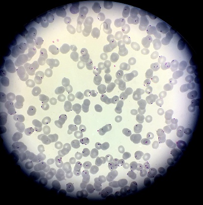
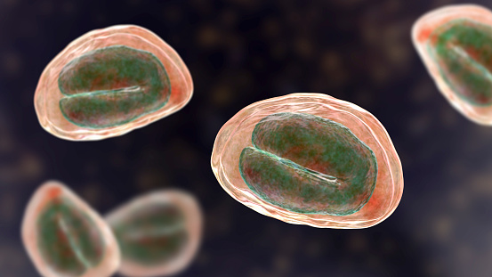
Free Images: "bestof:Plasmodium falciparum (malaria) parasite in blood.jpg Ring forms of the Plasmodium falciparum malaria parasite inside red blood cells Microscope image using"
female-mosquito-anopheles-insect-1332276.jpg
mosquito-insect-sucker-blood-pest-26057.jpg
mosquito-malaria-plaque-disease-1016254.jpg
mosquitoe-mosquito-malaria-gnat-1548976.jpg
mosquito-malaria-gnat-bite-insect-1548948.jpg
mosquitoe-mosquito-malaria-gnat-1548975.jpg
mosquito-malaria-gnat-bite-insect-1548947.jpg
mosquito-malaria-gnat-bite-insect-1548946.jpg
mosquito-insect-winged-insect-bug-1421757.jpg
insect-macro-wings-magnification-1529383.jpg
mosquito-insect-winged-insect-bug-1489973.jpg
animal-biology-bite-black-blood-931570.jpg
1._Healthy_red_blood_cells.jpg
bee_small_red_tip,_f,_chile,_side_2014-08-08-17.43.13_ZS_PMax.jpg
bee_small_red_tip,_f,_chile,_face_2014-08-08-17.31.24_ZS_PMax.jpg
bee_small_red_tip,_f,_chile,_angle_2014-08-08-17.22.49_ZS_PMax.jpg
3._Red_blood_cells_showing_signs_of_yeast.jpg
blood-cells-cells-human-543484.jpg
blood-bags-red-red-blood-cells-91169.jpg
blood-bags-red-red-blood-cells-91170.jpg
blood-bags-red-red-blood-cells-91171.jpg
bug-mosquito-fly-insect-pest-774506.jpg
2._Unhealthy_red_blood_cells.jpg
mosquito-bug-insect-pest-disease-19487.jpg
Closeup-Of-A-Mosquito-Feeding-On-Blood.jpg
mosquito-macro-insect-bug-animal-719613.jpg
bee-insect-small-pest-bug-450432.jpg
bat-tick-tick-ventral-insect-541938.jpg
mosquito-insect-small-pest-bug-450394.jpg
mosquito-insect-fly-blood-eating-387707.jpg
boxing-ring-boxers-fight-fighting-100733.jpg
agar-breeding-ground-red-60571.jpg
tick-wood-tick-blood-sucker-hand-482613.jpg
Dermacentor_variabilis,_U,_Back,_MD,_Beltsville_2013-07-08-19.15.11_ZS_PMax.jpg
bacteria-electron-microscope-stained-808158.jpg
bacteria-electron-microscope-811861.jpg
image-painting-digital-painting-1535548.jpg
Epeolus_lectoides,_m,_face,_Dorchester_Co,_MD_2016-01-07-13.17.jpg
Epeolus_lectoides,_m,_back,_Dorchester_Co,_MD_2016-01-07-12.55.jpg
Pasites_maculatus,_f,_pakistan,_back_2014-11-02-01.39.25_ZS_PMax.jpg
Pasites_maculatus,_f,_pakistan,_side_2014-11-02-01.50.40_ZS_PMax.jpg
Pasites_maculatus,_f,_pakistan,_face_2014-11-02-01.46.02_ZS_PMax.jpg
Harmful_Water_Organisms.jpg
Triepeolus_lunatus,_f,_right_side,_Charleston_Co.,_SC_2016-09-08-18.37.jpg
bee_bright_geeen,_m,_argentina,_angle_2014-08-07-16.59.52_ZS_PMax.jpg
bee_bright_geeen,_m,_argentina,_face2_2014-08-07-17.21.49_ZS_PMax.jpg
bee_bright_geeen,_m,_argentina,_side_2014-08-07-17.43.37_ZS_PMax.jpg
Nomada_rubicunda,_f,_face,_Minnesota_2016-04-16-02.02.jpg
Triepeolus_lunatus,_f,_back,_Charleston_Co.,_SC_2016-09-08-18.14.jpg
Triepeolus_lunatus,_f,_face,_Charleston_Co.,_SC_2016-09-08-18.23.jpg
Paranomada_velutina,_m,_arizona,_face_2014-08-08-14.19.04_ZS_PMax.jpg
Nomada_rubicunda,_f,_right_side,_Minnesota_2016-04-16-02.16.jpg
Nomada_rubicunda,_f,_back,_Minnesota_2016-04-16-01.42.jpg
Physocephala_tibialis,_back,_scarpulla_,_virginia_2014-07-09-18.06.jpg
Lord_Byron_-_Childe_Harold's_Pilgimage_-_Dugdale_edition.jpg
Paranomada_velutina,_m,_arizona,_angle_2014-08-08-14.11.49_ZS_PMax.jpg
paranomada_velutina,_f,_arizona,_face_2014-08-09-15.30.33_ZS_PMax.jpg
paranomada_velutina,_f,_arizona,_angle_2014-08-09-15.21.39_ZS_PMax.jpg
Paranomada_velutina,_m,_arizona,_side_2014-08-08-14.29.42_ZS_PMax.jpg
Gazania_krebsiana.jpg
Physocephala_tibialis,_face,_scarpulla,_virginia_2014-07-09-18.19.jpg
Physocephala_tibialis,_side,_scarpulla,_vriginia_2014-07-09-18.25.jpg
modesta,_male,_face_2012-07-16-18.19.jpg
Nomada_rubicunda,_m,_back,_Worchester_Co,_MD_2016-02-18-10.53.jpg
Nomada_rubicunda,_m,_side,_Worchester_Co,_MD_2016-02-18-12.01.jpg
Nomada_rubicunda,_m,_face,_Worchester_Co,_MD_2016-02-18-11.26.jpg
modesta,_male,_underside_of_abdomen_2012-07-16-18.37.jpg
Andrena_nivalis,_stylopid,_U,_Cocos_Co.,_NH,_back_2015-07-13-15.57.01_ZS_PMax.jpg
Triepeolus_distinctus,_m,_anne_arundel_co,_md,_back_2016-08-31-17.40.jpg
Villarceau circles.gif
Pieter_Brueghel_the_Elder_-_The_Dutch_Proverbs_-_Google_Art_Project.jpg
Osmotic pressure on blood cells diagram.svg
Osmotic pressure on blood cells diagram (zh-cn).svg
Osmotic pressure on blood cells diagram pl.svg
Osmotic pressure on blood cells diagram-es.svg
Osmotic pressure on blood cells diagram-ru.svg
Osmotic pressure on blood cells diagram-lv.svg
bacteria-bacterial-species-imitation-108897.jpg
bacteria-bacterial-species-imitation-108898.jpg
bacteria-bacterial-species-imitation-108896.jpg
bacteria-bacterial-species-imitation-108895.jpg
Design_Analysis_of_the_Lockheed_PV-1_Ventura.png
Vulva symbols.svg
portugues-sexo_alemão-sex_ingles-sex_africano-seks_russo-секс_albanes-sex_chines-性別_bulgaro-секс_croata-spol_gales-rhyw_coreano-섹스_frances-sexe_japones-セックス_filandes-sukupuolesta_arabe-الجنس_prontos_ai_tens_em_15_linguas_a_p.jpg
Symptoms of Raw fish infection.svg
Stem cell treatments.svg
BlankMap-World6.svg
Design_Analysis_of_the_F6F-5_Hellcat.png
red-blood-cells-red-mentally-mental-562852.jpg
blood-blood-plasma-red-blood-cells-75302.jpg
blood-blood-plasma-red-blood-cells-75301.jpg
mosquito-insect-bug-pest-malaria-37881.svg
Red-blood-cell illust.svg
cells-cell-structure-organism-blood-75305.jpg
Osmotic pressure on blood cells diagram-sk.svg
mosquito-bug-wings-sucker-insect-48313.svg
Terms of Use   Search of the Day