Click Here for More Images from iStock- 15% off with coupon 15FREEIMAGES 
GABA B receptors  are G protein-coupled receptors, also called metabotropic receptors.  Binding of  an agonist (baclofen, red) leads to a G-protein coupled C-AMP signaling pathway. Source: PDB entries 7eb2, 6r3q,.
Endosomes - 3d rendered image of Endosomes are membrane-bound organelles. They are parts of endocytic membrane transport pathway.  Endomembrane system in cells. Bilayer Lipid Membrane. Abstract medical illustration. Microscopic view. Microbiologic research concept.
Proteasomes are molecular machines for breaking down proteins called proteolysis, only target proteins that have been marked for destruction called ubiquitin to the target protein, 3d rendering
A ball and stick model of Human Insulin. Isolated on black.
3D cartoon and Gaussian surface models, chain id color scheme, PDB 7t3a, white background
Inside protein molecule
Covid-19 Coronavirus Variants Omicron Delta Gamma Beta Alpha Pandemic Virus Disease Mutation
3D rendering of alpha-synuclein protein aggregate
The glucagon like peptide-1 receptor (GLPr) is a G proteincoupled receptor (GPCR) that mediates the action of GLP-1, a peptide hormone. The activated receptor  has a strong effect  on the management of type 2 diabetes mellitus and obesity, including glucose homeostasis and regulation of gastric motility and food intake.
Endosomes - 3d rendered image of Endosomes are membrane-bound organelles. They are parts of endocytic membrane transport pathway.  Endomembrane system in cells. Bilayer Lipid Membrane. Abstract medical illustration. Microscopic view. Microbiologic research concept.
Intracellular transport, kinesin motor proteins, orange, transport molecules moving across microtubules, 3D illustration
3D Rendered Illustration, visualization of Neurons firing neurotransmitters in the synaptic gap.
3D cartoon and Gaussian surface models, PDB 2j0D, white background.
Cells in the body examined by endoscope
A ball and stick + ribbons model of beta-secretase - a protein that is important in forming healthy peripheral nerve cells, but is also involved in the onset of Alzheimer's disease.
3D rendering of forming human apoptosome molecule contains seven Apaf-1 molecules symmetrically arranged in a wheel-shaped structure to form a central hub
p53 (blue) tagged with ubiquitin (yellow) is degraded into small peptides by a proteasome (violett). p53 prevents cancer formation and acts as a guardian of the genome. Mutations in the p53 gene contribute to about half of the cases of human cancer.
Endosomes - 3d rendered image of Endosomes are membrane-bound organelles. They are parts of endocytic membrane transport pathway.  Endomembrane system in cells. Bilayer Lipid Membrane. Abstract medical illustration. Microscopic view. Microbiologic research concept.
3d rendered Gastric inhibitory polypeptide (GIP, glucose-dependent insulinotropic peptide) endocrine protein hormone, 3D illustration
What micro plastic is made of - Chemical ingredients.\nClose up of microplastic on finger concept of water pollution and global warming
3D cartoon and Gaussian surface models, chain id color scheme, PDB 3omv, white background
Digital background depicting innovative technologies in (AI) artificial systems, neural interfaces and internet machine learning technologies
NSCLC is any type of epithelial lung cancer other than small cell lung cancer SCLC. The most common types of NSCLC are squamous cell carcinoma, large cell carcinoma, and adenocarcinoma, but there are several other types that occur less frequently, and all types can occur in unusual histologic variants.
Antibodies in vivo. Antibody-based therapies have recently been studied. They ar\ne often the focus of attention in chemistry topics. It is a topic that should be developed very much. They are not generated by AI.
From left to right: the NMDA and  AMPA receptors transport calcium cations into neurons after being activated by the neurotransmitter glutamate and  the GABA receptor (right) transport chloride anions after the activation by gamma aminobutyrate. PDB entries: 6wht,  3kg2, 6d6u
Endosomes - 3d rendered image of Endosomes are membrane-bound organelles. They are parts of endocytic membrane transport pathway.  Endomembrane system in cells. Bilayer Lipid Membrane. Abstract medical illustration. Microscopic view. Microbiologic research concept.
Epstein-Barr virus internal structure. 3d illustration isolated in white
Cell Membrane Phospholipid Structure,Phospholipid bilayer of the cell membrane,Lipid bilayer
Cell membrane cholesterol is shown in pink. 3D cartoon and Gaussian surface models, PDB 7rpk, white background.
Myxoid liposarcoma.  Myxoid/round cell liposarcoma, or MRCLS, is one of several types of liposarcoma. Liposarcoma is a rare cancer that grows in the cells that store fat in the body. MRCLS usually grows in the arms and legs. These tumors grow slowly, and they can spread to other parts of the body
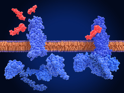
Free Images: "bestof:PDB 1osx EBI.jpg Small proteins TNF receptor-like TNF receptor-like BAFF receptor-like Tumor necrosis factor receptor superfamily member 13c BAFF-R Br3 EBI"
antenna-receptor-space-pot-377594.jpg
radio-old-vintage-receptor-1227044.jpg
aerials-antenna-telecommunications-1471443.jpg
phone-phone-booth-communication-1344294.jpg
antenna-space-pot-star-cosmos-947821.jpg
antenna-space-pot-star-cosmos-947824.jpg
radio-old-vintage-handy-old-radio-1284961.jpg
old-radio-old-radio-transistor-1528083.jpg
radio-old-old-radio-transistor-1528088.jpg
radio-in-september-walkie-talkie-1224724.jpg
radio-set-radio-communication-1224721.jpg
radio-in-september-walkie-talkie-1224722.jpg
radio-in-september-walkie-talkie-1224731.jpg
Steatosis.svg
CD8 receptor.svg
EGFR signaling pathway.svg
EGFR signaling pathway (zh-cn).svg
5HT1A-schema.svg
Afatinib mechanism.svg
EGFR signaling pathway de.svg
Insulin glucose metabolism ZP.svg
Insulin glucose metabolism ZP-ca.svg
Human genome by functions.svg
Notable mutations.svg
Bisphenol A diglycidyl ether 200.svg
Monoamine receptor tree.svg
Acetylcholine nicotinic receptor binding.svg
Sweet receptor + glucose.svg
Fc receptor schematic.svg
Decamethonium binding acth-receptor.svg
ZK 216348 structure.svg
Trifluoroisopropanolamine SEGRA.svg
RU 24858 skeletal.svg
Protein 7 domen.svg
MK-5932 skeletal.svg
Epelsiban structure.svg
A 276575 structure.svg
Lafutidine.svg
CP154526.svg
ZM241385.svg
Octahydrophenanthrene-2,7-diol SEGRA.svg
OUP-16.svg
CX-516-2D-skeletal.svg
CPCCOEt.svg
Diphenidine.svg
Selurampanel.svg
Fimasartan skeletal.svg
Etacstil skeletal.svg
Sumatriptan binding site.svg
Alpha-Methylhistamine.svg
Aptiganel.svg
Oxiracetam.svg
Zibotentan.svg
nerve-cell-neuron-biology-science-145909.svg
NEFA.svg
Mozavaptan structure.svg
Semnalizare engl.svg
8-OH-DPAT.svg
DCKA.svg
Kynurenic acid.svg
Fananserin.svg
Dextrorphan.svg
Remacemide.svg
Niaprazine.svg
Ciproxifan.svg
Metomidate.svg
Alnespirone.svg
Zalospirone.svg
Deramciclane.svg
Propoxate.svg
Lasmiditan skeletal.svg
ORs and GP.svg
Velusetrag.svg
Idoxifene.svg
McN5652.svg
Milacemide.svg
Levormeloxifene.svg
Nitrosoglutathione.svg
WAY-161503.svg
PRX-03140.svg
EPI-001.svg
Acolbifene.svg
Filorexant.svg
Piromelatine.svg
Licostinel.svg
Raseglurant.svg
Pipoxizine.svg
Dasolampanel.svg
Fanapanel.svg
Becampanel.svg
Zonampanel.svg
Merotocin.svg
Tetrahydrochrysene.svg
Broparestrol.svg
Estrobin.svg
Macitentan skeletal.svg
ACT-132577 skeletal.svg
Etacstil.svg
Endoxifen.svg
Eprosartan.svg
Terms of Use   Search of the Day