Click Here for More Images from iStock- 15% off with coupon 15FREEIMAGES 
Mosquito Borne Virus risk like Zika  Dengue and Malaria spread through Aedes and Anopheles mosquitoes causing infectious diseases with 3D illustration elements.
Bacteria Aggregatibacter, 3D illustration. Aggregatibacter aphrophilus and A. actinomycetemcomitans, Gram-negative bacteria, part of the normal flora of the mouth and throat, also cause endocarditis
Mosquito under microscope
Microalgae under microscopic view, green algae, cyanobacteria, phytoplankton, diatom, algae mix collage background
Photomicrograph of filamentous blue-green algae, Oscillatoria species. Each band along the stand is one cell. This filament is very short probably having recently parted from a longer one. Filaments are typically many times longer. This algae is mobile, it moves and twists through the water. San Francisco Bay, California, USA. Live specimen. Wet mount, 40X objective, transmitted brightfield illumination. Note - motion blur of live specimen, very shallow depth of field, chromatic aberration and uneven focus are inherent in light microscopy.
Sciaroidea insect Fossil inside Burmese Amber of Cenomanian Era, 100 million years ago, from the state of Myanmar
Leiden leaf c.s. under microscope
Epidermis. Electron microscope micrograph showing a keratinocyte of spinous layer. The epithelial cell has a polygonal shape, central nucleus with nucleolus, cytoplasm full of keratin filament bundles, and numerous dark desmosomes crossing the intercellular spaces.
Particles, medical concept
Scanning electron micrograph, black&white
Burkitt lymphoma is a form of non-Hodgkin's lymphoma in which cancer starts in immune cells called B-cells. Recognized as a fast growing human tumor, Burkitt lymphoma is associated with impaired immunity and is rapidly fatal if left untreated.
Diatoms, algae under microscopic view, phytoplankton, fossils, silica, golden yellow algae
plant Zea Stem C.S. under light mircoscope with white background
Mosquito on human skin sucking blood diagonal view background 3d rendering
Mosquito under a microscope
Mosquito trying to bite through a gray matter
Natural agate surfaces, suitable for backgrounds and wallpapers.
Bacteria Aggregatibacter, 3D illustration. Aggregatibacter aphrophilus and A. actinomycetemcomitans, Gram-negative bacteria, part of the normal flora of the mouth and throat, also cause endocarditis
front view of a damselfly
Euglena is a genus of single cell flagellate eukaryotes under microscopic view for study.
Escherichia Coli (E. Coli.)
insect larva ( Orthocladiinae - order diptera ) under the microscope - light microscope x200 magnification
Portrait of Blow Fly on blue
fish skin under light microscope with white background
Extreme macro photography of a mosquito. Multiple images taken using focus rail and stacked using Helicon focus software.
Microscope photo of a sambucus stem with parenchyma cells.
Pheochromocytoma, Site: Adrenal gland
Eggs of helminths Enterobius vermicularis containing worm larva. Threadworm which cause enterobiasis, 3D illustration
Simuliidae, black fly (Diptera) in Baltic amber, Eocene, approximately 56 - 34 million years ago. Image taken with extreme macro and focus stacking technique.
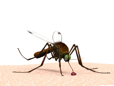
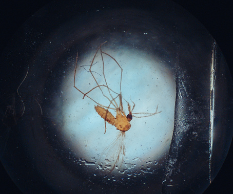
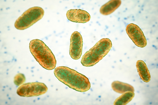
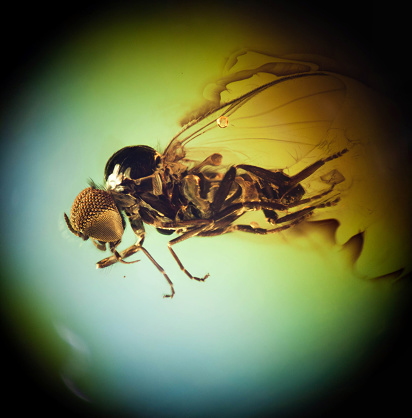
Free Images: "bestof:Malaria Plasmodium gallinaceum.jpg Scanning electron micrograph of Plasmodium gallinaceum invading mosquito midgut Categories Research in NIH Labs and Clinics"
female-mosquito-anopheles-insect-1332276.jpg
Ebola_Virus_Particles.jpg
Malaria_Plasmodium_gallinaceum.jpg
HeLa-IV.jpg
MRSA_dead_neutrophil.jpg
HIV_H9_T-cell_II.jpg
White_blood_cell_with_MRSA.jpg
Dendritic_cell_revealed.jpg
Surface_of_HIV_infected_microphage.jpg
SIV_virion_in_3D.jpg
Malaria_LifeCycle(French_version).GIF
Embryonic_smooth_muscle_cell.jpg
T_cell_and_virus.jpg
HIV_spike_in_complex.jpg
Hemozoin crystals.jpg
Malaria-infected_Red_Blood_Cell.jpg
Skin surface (human).jpg
HIV_H9_T-cell.jpg
MRSA,_Ingestion_by_Neutrophil.jpg
Salmonella_NIH.jpg
Brain_Scan_-_Methamphetamine.jpg
Balance_testing.jpg
Hypothalamus_calretinin_nuclei.jpg
Habenula_mouse.jpg
Drug_Exposure_Brain.jpg
Craniofacial_mouse_embryo.jpg
3D_pyruvate_dehydrogenase_complex.jpg
Drug_Brain_Activity.jpg
Bowel_vasculature_-_1.jpg
Bowel_vasculature.jpg
Hippocampus_mossy_fiber_interneuron.jpg
Cerebellum_Purkinje_nuclei.jpg
Piriform_cortex_of_a_mouse.jpg
Dentate_gyrus_II.jpg
CA1_hippocampus.jpg
Transgenic_mouse_hippocamus.jpg
Sagittal_mouse_cannabinoid_receptor_nuclei.jpg
Dentate_gyrus.jpg
Confocal_image_of_spiny_neuron_-_1.jpg
Confocal_image_of_spiny_neuron.jpg
Mouse_Tractography.jpg
Stereocilia_of_frog_inner_ear.01.jpg
Plasmodium.jpg
Nanoparticles; scanning electron micrograph (5887833046).jpg
"Life_History_of_the_Malaria_Parasite_(Plasmodium_Vivax)_in_man_and_the_Anopheles_Mosquito"_-_NARA_-_514892.tif
"Life_History_of_the_Malaria_Parasite_(Plasmodium_Vivax)_in_man_and_the_Anopheles_Mosquito"_-_NARA_-_514892.jpg
Yersinia_pestis.jpg
HeLa-V.jpg
Plasmodium_falciparum_rings_form_parasites4885_lores.jpg
Electronmicroscopynhlbi.jpg
Neutrophil_MRSA_I.jpg
Streptococcus_pyogenese_Bacteria.jpg
Schistosoma mansoni2.jpg
Nanoknife.jpg
Human_neutrophil_ingesting_MRSA.jpg
Yersinia_pestis_Bacteria.jpg
Aspergillus.gif
Anthrax_color_enhanced_micrograph.JPG
Neutrophil_MRSA_II.jpg
MRSA_SEM_7821_lores.jpg
MalariacycleBig.jpg
Malaria_parasite_life_cycle-NIAID.jpg
Red_blood_cell_infected_with_malaria_parasites-NIAID_RML.jpg
Tropeninstitut berlin.jpg
Microblind.JPG
Gametocytes_plasmodium_5952_lores.jpg
Stereocilia.jpg
Macrogametocyte_of_the_parasite_Plasmodium_falciparum_PHIL_2701_lores.jpg
Microgametocyte_of_the_parasite_Plasmodium_vivax_PHIL_2727_lores.jpg
Cairns AWM119073.jpg
Zbar, berton (1).jpg
NIH clinical center (4).jpg
Macrophages in the brain before and after in vitro infecti.jpg
Fluorescent image fibroblast.jpg
Hhv-6 (1).jpg
Terms of Use   Search of the Day