Click Here for More Images from iStock- 15% off with coupon 15FREEIMAGES 
Human anatomy detail of shoulder. Muscle, bone structure, arteries. On plain studio background. Professional lighting.
An acupuncture model of the male human torso depicting the acupoints and meridiens used in Traditional Chinese Medicine and massage therapy
Man's upper body muscles frontal anterior view labeled on a white background.
MRI scanning of the human body skeletal, muscular and nervous systems with abdomen region view. / You can see the animation movie of this image from my iStock video portfolio. Video number:
The posterior sacroiliac ligament is a compound ligament composed of three distinct bands 3d illustration
Cervical Vertebrae Bridge X-ray Film
Shoulder or glenohumeral joint close-up 3D rendering illustration isolated on white with copy space. Human skeleton anatomy, medical diagram, osteology, skeletal system, science, biology concepts.
Computational tomography image of the abdominal and pelvic regions.
Acupuncture model with chinese
Gluteus Medius Muscle anatomy for medical concept 3D illustration
Human anatomy of male forearm Extensor Digitorum muscles isolated within the skeletal system bones in a rear view on a black background.
Xray view of a patient with an injured spine and suffering. / You can see the animation movie of this image from my iStock video portfolio. Video number: 1453180480
medically accurate muscle illustration of the rhomboid major
X-ray of female hips, pelvis and spine
Human lumbar vertebrae with spinal cord and nerve isolated on white background 3D rendering illustration. Blank anatomical chart. Anatomy, medical and healthcare, science, medicine concepts.
Film x-ray human's pelvis
The lumbar vertebrae are, in human anatomy, the five vertebrae between the rib cage and the pelvis.
waist pain three Dimensional
The vertebral column surrounds the spinal cord which travels within the spinal canal, formed from a central hole within each vertebra
Computed MRI image of Prostate Cancer.
Labeled human anatomy of male forearm Exterior Carpi Ulnaris muscles isolated within the skeletal system bones in a rear view on a white background.
Digital medical illustration: Human spine featuring vertebrae (cervical (C1-C7), thoracic (T1-T12) and lumbar (L1-L5)) vertebrae, discs and pelvis.
Femur bones in red color with body 3D rendering illustration isolated on white with copy space. Human skeleton and leg anatomy, medical diagram, osteology, skeletal system, science concepts.
Computer futuristic digital HUD software interface with a system for scanning the human body and transmitting images and signals in real time. Within one complex, both a scanner and a digital video recorder are used.
3D Illustration Concept of Human Skeleton System Pectoral (Shoulder) Girdle Bone Joints Anatomy
Backlit image of fake bones made of resin.
Human Anatomy,  Limbs and hip skeletal, muscular and cardiovascular systems, with sub layers muscles. front anche rear views, on white background. 3d Illustration
Xray of ones manhood.
The posterior sacroiliac ligament is a compound ligament composed of three distinct bands 3d illustration
Cat x-ray image
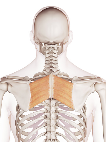
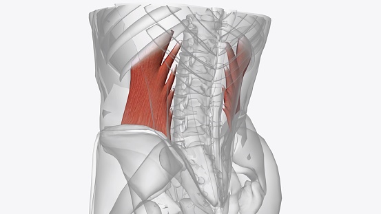
Free Images: "bestof:Latissimus dorsi.PNG Muscles connecting the upper extremity to the vertebral column Image Gray409 png 2007-10-10 User Mikael Häggström Gray's Anatomy plate"
Naked human male body front anterior.png
Human anatomy.jpg
Anatomía humana.png
Human anatomy ru 2.jpg
Naked human male body front anterior (annotated-en).png
Persons.jpg
Sarychev Volcano edit.jpg
Lewis_Hine_Power_house_mechanic_working_on_steam_pump.jpg
Haeckel_Batrachia.jpg
Prepatellar_bursa_(vector).svg
Maxillary_artery_branches.svg
Surface projections of the organs of the trunk.svg
Axillary_lines.svg
Placenta.svg
Glucose metabolism.svg
Smallpox versus chickenpox english plain.svg
Gray696.svg
Diagram of influenza infection symptoms EN.svg
Axial skeleton diagram blank.svg
Human body silhouette.svg
Surface anatomy of the heart.svg
Placenta_ja.svg
Placenta_ko.svg
Placenta_de.svg
Placenta_ar.svg
Man shadow anatomy.svg
Internal organs.svg
Gray1220.svg
En-elbow joint.svg
Female shadow anatomy.svg
Gray1142.png
Muscles_of_the_male_perineum-Gray406.png
Symptoms of swine flu-es.svg
Pioneer plaque man upper body as diagram template.svg
Pioneer plaque man upper body as diagram template (alternative).svg
Female shadow anatomy without labels.svg
Gray1235_(English).svg
Gray826and831.svg
Symptoms of swine flu-nl.svg
Surface projections of the organs of the trunk - without labels.svg
Gray1167.svg
Gray1167_fi.svg
Smallpox versus chickenpox neutral plain.svg
Smallpox versus chickenpox latin plain.svg
Dermatomes and cutaneous nerves - posterior.svg
Vaiolo VS Varicella.svg
Gray1167_de.svg
Gray814.svg
Gray1220-es.svg
Gray1236_pl.svg
Gray1235_pl.svg
Gray1167 es.svg
Gray728.svg
Gray726-Brodman.svg
Gray726-Brodman-prefrontal.svg
Gray1095_-_pl.svg
Gray38.svg
Gray812and814.svg
Gray1236.svg
Gray1235.svg
Gray67.svg
Gray512.svg
Gray834 pl.svg
Gray814_pl.svg
Brain_diagram_ja.svg
Brain_diagram_pl.svg
Heart sounds auscultation areas.svg
Brain_diagram_without_text.svg
Gray1095.svg
Effects of sleep deprivation.svg
PD Diagram of swine flu symptoms EN.svg
Diagram of swine influenza symptoms EN.svg
Design_Analysis_of_the_Lockheed_PV-1_Ventura.png
Design_Analysis_of_the_P-38_Lightning.png
Dermatomes and cutaneous nerves - anterior.svg
Tyrosine hydroxylase (zh-cn).svg
Lymphocyte activation.svg
Liberty drawing.svg
Cystic fibrosis manifestations.svg
Arterial system.svg
Symptoms of multiple sclerosis es.svg
Gray424.svg
Gray1154.png
Gray588.png
Gray1155.png
Lymphocyte activation simple.svg
Intermediate filament sr.svg
Humerus.svg
Paranasal sinuses transparent.svg
Симптомы эболы (векторная версия).svg
Gray1155-IT.png
Human skeleton front - no labels.svg
Side effects of aspirin.svg
Symptoms of aspirin overdose.svg
Symptoms of bubonic plague.svg
Symptoms of pneumonic plague.svg
Pioneer plaque man as diagram template.svg
Symptoms of SLE.svg
Human skeleton (svg template).svg
Drumstick anatomy.svg
Terms of Use   Search of the Day