Click Here for More Images from iStock- 15% off with coupon 15FREEIMAGES 
Pine mature wood cross section. Light microscope slide with microsection of an evergreen conifer in the genus Pinus. Plant anatomy. Biology. Photo.
plant Tilia Stem C.S. under light mircoscope with white background
The study of plant tissues under the microscope in the laboratory.
Natural agate surfaces, suitable for backgrounds and wallpapers.
Stratified flat epithelium under microscope
Particles, medical concept
Cells isolated on white background
corn stem micrograph with dye
Virus 3d model set. Covid-19 germs, fungi, bacteria objects. Graphic from microscopic zoom in lab for learning science medical, biology, virology on white background. Clipping path. 3D Illustration.
Focal nodular hyperplasia (FNH) is a benign liver lesion that is composed of a proliferation of hyperplastic hepatocytes surrounding a central stellate scar. Typically, FNH is a solitary lesion that is more commonly seen in women.
Blue colored stem cells of a lentil plant under the microscope.
Quantum Gravity 3d rendered image of a comprehensive exploration of Quantum Gravity theories. String theory, loop quantum gravity (LOG), causal dynamical triangulation, twistor theory, gravitational waves, spacetime, gravity waves, quantum mechanics, quantum entanglement concepts.
Leiden leaf c.s. under microscope
Abstract particles
Transmission electron micrograph (TEM) showing a striated skeletal muscle cell in cross section. Among myofibrils (sectioned at the band A level), mitochondria and sarcoplasmic reticulum can be seen.
Slice of natural crystal for patterns and textures use with high resolution.
Photomicrograph of filamentous blue-green algae, Oscillatoria species. Each band along the stand is one cell. This filament is very short probably having recently parted from a longer one. Filaments are typically many times longer. This algae is mobile, it moves and twists through the water. San Francisco Bay, California, USA. Live specimen. Wet mount, 40X objective, transmitted brightfield illumination. Note - motion blur of live specimen, very shallow depth of field, chromatic aberration and uneven focus are inherent in light microscopy.
Host cells with spores (mold) are inside wood under the microscope for education.
Microscopic fungi Malassezia furfur, showing yeast cells and hyphae. dermatophytes, Nail scraping or skin scraping for fungus test in microbiology laboratory.
Blastomycosis is a fungal infection caused by inhaling Blastomyces dermatitidis spores. If it involves only the lungs, it is called pulmonary blastomycosis. Only about half of people with the disease have symptoms, which can include fever, cough, night sweats, muscle pains, weight loss, chest pain, and feeling tired.
Microalgae under microscopic view, green algae, cyanobacteria, phytoplankton, diatom, algae mix collage background
Epidermis. Electron microscope micrograph showing a keratinocyte of spinous layer. The epithelial cell has a polygonal shape, central nucleus with nucleolus, cytoplasm full of keratin filament bundles, and numerous dark desmosomes crossing the intercellular spaces.
Digital Dispersed corona virus on petri dish. Animation of multiplication of CORONA VIRUS COVID-19
PAPS Smear study of a young women under microscopy showing Atrophic changes in uterus which is unusual at young age.
Endosomes - 3d rendered image of Endosomes are membrane-bound organelles. They are parts of endocytic membrane transport pathway.  Endomembrane system in cells. Bilayer Lipid Membrane. Abstract medical illustration. Microscopic view. Microbiologic research concept.
Microscope photo of a sambucus stem with parenchyma cells.
Burkitt lymphoma is a form of non-Hodgkin's lymphoma in which cancer starts in immune cells called B-cells. Recognized as a fast growing human tumor, Burkitt lymphoma is associated with impaired immunity and is rapidly fatal if left untreated.
Paramecium is a genus of unicellular ciliated protozoa,  Paramecia are widespread in freshwater, brackish, and marine environments and are often very abundant in stagnant basins and ponds.
Curly marble stone with fashion veins.
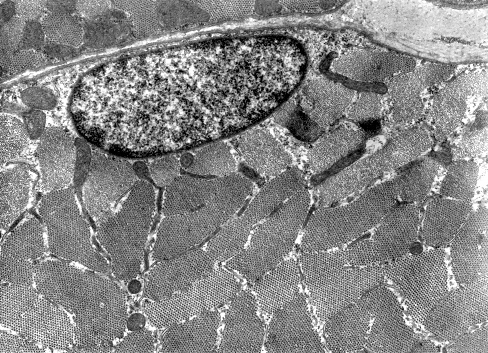
Free Images: "bestof:Hemozoin crystals.jpg en Electron micrograph of crystals of hemozoin isolated from the malaria parasite Plasmodium falciparum Magnified 68 490 times Original"
crystals-ice-crystal-snow-ice-67792.jpg
ice-crystal-crystals-snow-ice-67709.jpg
snow-crystals-ice-ice-crystal-67708.jpg
Pieter_Brueghel_the_Elder_-_The_Dutch_Proverbs_-_Google_Art_Project.jpg
Hexagonal Ice Crystals.svg
Diffusion lacunaire.svg
Atom1.svg
Diffusion interstitielle.svg
Edge dislocation 2.svg
Lacune cristal 2d.svg
Solution solide substitution insertion 2d.svg
Defaut antiphase cristal 2d.svg
Defaut frenkel cristal 2d.svg
Defaut schottky cristal 2d.svg
Zuber & Cie - Sidewall - Google Art Project (6818798).jpg
Design_Analysis_of_the_P-38_Lightning.png
Hemozoin crystals.jpg
P.falciparum hemozoin.jpg
Birefringent P.falciparum Pigment.jpg
Birefringence of malaria pigment.jpg
P.falciparum schizont.jpg
Plasmodium.jpg
Hemoglobinopathies and the pathogenesis of Plasmodium falciparum malaria - Journal.ppat.1003327.g001.png
Hemoglobinopathies-Slicing-the-Gordian-Knot-of-Plasmodium-falciparum-Malaria-Pathogenesis-ppat.1003327.g001.jpg
Spermatozoa-human-3140x.jpg
Spermatozoa-human-1000x.jpg
Red_blood_cell_infected_with_malaria_parasites-NIAID_RML.jpg
Nanowire; Practical Oscillators (5941044200).jpg
Schistosoma 20041-300.jpg
Gradient centrifugation of infected Red Blood Cells.jpg
The_Quarterly_journal_of_the_Geological_Society_of_London_(13937158391).jpg
Plasmodium falciparum (malaria) parasite in blood.jpg
Hemozoin structure.png
Macrogametocyte_of_the_parasite_Plasmodium_falciparum_PHIL_2701_lores.jpg
Malaria_Plasmodium_gallinaceum.jpg
Plasmodium_falciparum_rings_form_parasites4885_lores.jpg
Malaria-infected_Red_Blood_Cell.jpg
P.falciparum Gelatine enrichment.jpg
Malaria_parasite_life_cycle-NIAID.jpg
Cubicpyrite.jpg
Plasmodium falciparum in Red Blood Cells.jpg
Glucose-6-Phosphate Dehydrogenase activity stain.jpg
Blood stages of P. falciparum.jpg
Blood clot in scanning electron microscopy.jpg
Malaria_lifecycle-CDC.gif
Synchronsed P falciparum cultures.jpg
Rubrene.jpg
Pentacene.jpg
Drug-heme interaction.png
Blood stages of P. falciparum over 48h.jpg
Electron_Micrograph_of_Flea_derivate.jpg
Layered Approach May Yield Stronger, More Successful Bone Implants (5880495301).jpg
VaccHeuser.jpg
The spatial distribution of Plasmodium vivax malaria endemicity map in 2010 globally.png
Malaria_LifeCycle(French_version).GIF
Cleavage-furrow.JPG
Cinnabarr.jpg
Filter paper 840 3x3 copy.jpg
TBuOH 1133.jpg
AceticAcid010.png
Plasmodium falciparum 02.jpg
LDH.PNG
Bt-toxin-crystals.jpg
Microblind.JPG
Rosa epicuticular wax stoma.jpg
HeuserNeuro.jpg
Mineral crystals, Bruce Museum of Arts and Science.jpg
MalariacycleBig.jpg
The_Oak_(Marshall_Ward)_Fig_17.jpg
10-0002-F1.png
Bentley snowflake micrograph no. 777.jpg
Footprint.jpg
Atom1.png
AB Crystal 1593.JPG
OakCreekBigPark air.jpg
Na3FeOx3 01.JPG
BritishTunicataPlate57.jpg
Parker Valley facing S.jpg
Chalcopyrite.owl.arp.600pix.jpg
A gossypii.jpg
BritishTunicataPlate59.jpg
Rydberg atom animation.gif
Snow-crystal photo-micrograph, made by W. A. Bentley, Nashville (Jericho), Vermont, c. 1890s - Fairbanks Museum and Planetarium - DSC04382.JPG
BritishTunicataPlate58.jpg
Moeraki_Boulders_at_Dawn.jpg
Edge dislocation.png
Rydberg atom movie small.gif
The_Quarterly_journal_of_the_Geological_Society_of_London_(12960401793).jpg
KomatiiteA2.jpg
Phase drift.png
Geomyces_destructans.gif
Defecttypes.png
TwistGeometry.jpg
Andrew parodi and friend lec at graduation from western oregon university.jpg
Cystinosis.png
KomatiiteCanada 682By512.jpg
KomatiiteA3.jpg
Hexagonal Depository of the Scriptures-Koyasan-Danjogaran.jpg
Illustrated_Life_Cycle_of_Echinostoma.gif
The_Quarterly_journal_of_the_Geological_Society_of_London_(12960682974).jpg
Terms of Use   Search of the Day