Click Here for More Images from iStock- 15% off with coupon 15FREEIMAGES 
Illustration showing chromosomes with highlighted telomeres
Abstract Genetics Disease - 3d rendered image.
Dna structure on science background. 3d illustration
Chromosome on white background. 3D render.
Licmophora sp. algae, marine and freshwater diatom under microscopic view. Genus of benthic, photosynthetic and epiphyte diatom
Photomicrograph of filamentous blue-green algae, Oscillatoria species. Each band along the stand is one cell. This filament is very short probably having recently parted from a longer one. Filaments are typically many times longer. This algae is mobile, it moves and twists through the water. San Francisco Bay, California, USA. Live specimen. Wet mount, 40X objective, transmitted brightfield illumination. Note - motion blur of live specimen, very shallow depth of field, chromatic aberration and uneven focus are inherent in light microscopy.
centropyxis aculeata (amoeboid organism) under the microscope - optical microscope x400 magnification
Abs Virus - 3d rendered image like colorized scanning electron micrograph. Scene showing virus (yellow)  emerging from infected cell.\nConcept of coronavirus that’s causing a global pandemic. Covid-19, Sars-cov-2 alpha, betta, gamma, delta, lambda variant, 2019-nCoV, Coronavirus.\nSEM, TEM, hologram view.
Euglena is a genus of single cell flagellate eukaryotes under microscopic view for study.
Fetus with dna high quality 3D rendering
Chromosomes 3d illustration
chromosome,Human genome concept
Chromosome isolated on white background. with depth of field effect, scientific concept. 3d illustration
DNA and biotechnology concept
Y Chromosome abstract scientific background. 3D illustration
Detailed Human Stem Cell
Chromosome with blue background, 3d rendering. Computer digital drawing.
Liver Fluke Section under light microscope with white background
Microalgae under microscopic view, green algae, cyanobacteria, phytoplankton, diatom, algae mix collage background
Genetics background. 3D render.
Freshwater Closterium algae (unicellular charophyte green algae) - optical microscope x200 magnification
Photomicrograph of Euglena species. Fresh water, California. Live specimen. Wet mount, 40X objective, transmitted bright field illumination.
The intermembrane transporter of the Gram-negative bacterium Acinetobacter baylyi transports a lipopolysaccharide (orange) through the inner membrane consuming ATP (red)
Textured cells under heavy magnification
DNA digital data storage is the process of encoding and decoding binary data to and from synthesized strands of DNA 3d rendered
plant Leaf of Nerium indicum T.S. under light mircoscope with white background
Salmonella are a bacteria responsible for food poisoning, 3d render of microorganism cousing gastrointestinal disease
Trematode parasite under a microscope
DNA molecules, structure of the genetic code, 3d rendering,conceptual image.
Immunoglobulin G (IgG) - 3d rendered image. Microbiology illustration  the structure of an antibody. The Y-shaped structure. IgG is the most common type of antibody. Hologram view. SEM.
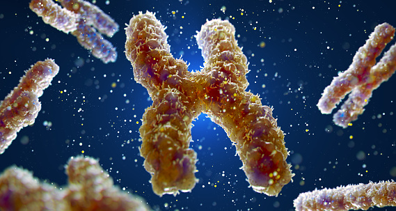
Free Images: "bestof:HLA-mini.png en Illustration of HLA-locus on chromosome 6 own Pdeitiker 08-07-2008 Human genes"
Albero Y-DNA.svg
HLA.svg
HLA-cs.svg
Atlas-BRCA1.svg
2009_H1N1_influenza_virus_genetic-num.svg
Vulva symbols.svg
HLA-mini.png
HLA-DQ locus.png
TMRCAs-compared.PNG
Xenoma3 miguelferig.jpg
HLA B^1501.png
HLA.jpg
DQ Illustration.PNG
Genelocation2.png
Human_chromosome_2_ideogram.png
Human chromosomesXXY02.png
PPHSimage01.jpg
Human_chromosome_11_ideogram.png
Xenoma4 miguelferig.jpg
Genomic_Context.PNG
KIAA0895Lneighborhood.JPG
Lactase Processing.png
DQ Illustration (rou).png
C11orf73aachangesperyeargraph.jpg
C11orf73identityvsdivergencegraph.jpg
A2-gliadin-33mer.png
Illustration HLA-A.png
KIAA1958 neighboring genes on both sides.jpg
Chromosome.gif
HLAn(ro).png
AACOX.png
Gray1211_(no_labels).png
Piskacek p53a.jpg
Piskacek p53b.jpg
5'UTR.png
IQGAP1.jpg
KIAA0895location.png
Tmem126B cartoon2.jpg
Allosteric.png
IQGAP1 pic2.jpg
Thalassemia_beta.jpg
Frecuencias en el chr22 (Dunham) (corregido).PNG
Early diversification.PNG
Tmem126B cartoon.large.jpg
Kit expression in hematopoietic cells.JPG
MSX1.jpg
Genes and bases on chromosomes.png
Human proteins - Conceptual translation of TMEM126B.png
3 Korean Samples.png
Scf in hsc niche.JPG
HLA-B.png
A1BGneighb.fcgi.png
Gliadin-immuno-innate.PNG
Protein digestion.PNG
2Nrule.PNG
A9-gliadin-innate.png
Normal Villus Illustration.png
HLA-DQ2.5-2.2 protein.png
Retrospective population bottleneck.PNG
HLA-DQ protein.PNG
Gene structure of the catalytic subunit isoforms of PKA.jpg
2cs4 PDB n-terminal domain chromosome 12 ORF 2.png
HLA-A2.png
Ube3a.jpg
Structure cornea.png
Structure lens.png
HLA-Cw4.png
A2-peptide.PNG
Paul Burridge Brachyury in E7.5.jpg
Huxley races.png
HLA-A1.png
Comparison of 3 by Y-DNA haplogroups in Northwestern Europe.png
Galten_Herreds_våben_1648.png
Settlement_in_the_East_Indies_WDL11282.png
The_Fight_Between_the_Sacrifice_and_He_Who_Sacrifices_WDL6757.png
The_Battle_of_Azcapotzalco_WDL6746.png
Camaxtli,_God_of_War_of_the_People_of_Tlaxcala_WDL6727.png
Tlacaxipehualiztli,_Festival_of_the_Flaying_of_Men,_the_Second_Month_of_the_Aztec_Solar_Calendar_WDL6735.png
Notes_on_Architectural_Perspective_WDL11281.png
Landscape_and_Notes_on_Shading_and_Perspective_WDL11283.png
Zeravshan_Okrug._Palanquins_of_Saint_Musa_Khan_Preserved_in_the_Mosque_of_Saint_Makhdum_Azam_in_the_Village_of_Dakhbit_WDL3896.png
Zeravshan_Okrug._Palanquins_of_Saint_Musa_Khan_Preserved_in_the_Mosque_of_Saint_Makhdum_Azam_in_the_Village_of_Dakhbit_WDL3897.png
Itzcóatl,_the_Fourth_Aztec_King_(Reigned_1427–40)_WDL6721.png
Tizoc,_the_Seventh_Aztec_King_(Reigned_1481–86)_WDL6722.png
Antiquities_of_Samarkand._Mausoleum_of_Khodzha_Abdu-Derun._General_View_of_the_Mausoleum_from_the_Southwest_WDL3734.png
Antiquities_of_Samarkand._Madrasah_of_Shaybani_Khan_and_Ruins_of_the_Tomb_of_the_Kuchkunji_Khans._View_of_the_Inner_Courtyard_and_Crypt_of_Shaybani_Khan_WDL3824.png
Antiquities_of_Samarkand._Mausoleum_of_the_Emir_Timur_Kuragan_(Gur-Emir)._Interior_of_the_Tomb._View_of_the_Family_Crypt_of_Tamerlane_WDL3574.png
Antiquities_of_Samarkand._Mausoleum_of_Emir_Timur_Kuragan_(Gur-Emir)._Plans,_Elevations,_and_Sections_WDL3575.png
Antiquities_of_Samarkand._Mausoleum_of_the_Emir_Timur_Kuragan_(Gur-Emir)._Section_of_Marble_Lattice-Work_Surrounding_the_Gravestone_WDL3713.png
Antiquities_of_Samarkand._Mausoleum_of_the_Emir_Timur_Kuragan_(Gur-Emir)._Section_of_Grave_Marker_WDL3714.png
Antiquities_of_Samarkand._Mausoleum_of_the_Emir_Timur_Kuragan_(Gur-Emir)._Part_of_the_Foundation_inside_the_Mausoleum_WDL3715.png
Antiquities_of_Samarkand._Mausoleum_of_the_Emir_Timur_Kuragan_(Gur-Emir)._Main_Door_on_the_Northern_Facade_WDL3717.png
Antiquities_of_Samarkand._Mausoleum_of_the_Emir_Timur_Kuragan_(Gur-Emir)._Column_Capital_and_Section_of_Detail_on_the_Northern_Facade_WDL3718.png
Antiquities_of_Samarkand._Mausoleum_of_the_Emir_Timur_Kuragan_(Gur-Emir)._Inscriptions_around_the_Frieze_of_the_Entire_Front_WDL3722.png
Antiquities_of_Samarkand._Mausoleum_of_the_Emir_Timur_Kuragan._View_of_the_Western_Facade_of_the_Mausoleum_WDL3731.png
Antiquities_of_Samarkand._Madrasah_of_Tillia_Kari._Inner_Courtyard_(Southern_Side)._Main_Niche_WDL3810.png
Antiquities_of_Samarkand._Mausoleum_of_the_Emir_Timur_Kuragan_(Gur-Emir)._Section_of_the_Foundation_inside_the_Tomb_WDL3573.png
Antiquities_of_Samarkand._Tomb_of_the_Emir_Timur_Kuragan_(Gur-Emir)._One_of_the_Doors_WDL3576.png
Antiquities_of_Samarkand._Mausoleum_of_the_Emir_Timur_Kuragan_(Gur-Emir)._Part_of_a_Corner_Column_of_the_Facade_WDL3716.png
Antiquities_of_Samarkand._Mausoleum_of_the_Emir_Timur_Kuragan_(Gur-Emir)._Inscription_on_One_of_the_Doors_WDL3719.png
Terms of Use   Search of the Day