Click Here for More Images from iStock- 15% off with coupon 15FREEIMAGES 
forming of underskin cellulite illustration
X-ray pictures or images of a woman's healthy breasts with no sign of breast cancer.
black armpit fat women
Serous carcinoma is the most common type of ovarian cancer, accounting for approximately 75% of epithelial ovarian cancers.
PAPS Smear study of a young women under microscopy showing Atrophic changes in uterus which is unusual at young age.
Fog or Smoke, From Above  textured
ultrasonography or ultrasound images of upper abdomen with image of gallbladder, liver, spleen, kidney, pancreas and aorta
Partial closeup of pink blanket
Wilms tumor, or nephroblastoma, light micrograph, photo under microscope
skin science 3d
3D Illustration Concept of Human Muscular System Arm Muscles Tricep Muscle Anatomy
Diffuse large B-cell lymphoma, or DLBCL, is a cancer that starts in white blood cells called lymphocytes.
Chaetoceros is the largest genus of marine planktonic diatoms, phytoplankton
abstract imagery of a fusion of colors and surfaces
TMJ: The temporomandibular joints. Healthy occlusion anatomy. Medically accurate 3D illustration of human teeth and dentures concept
Liver Fluke Section under light microscope with white background
Xray of ones manhood.
pork stomach on white background,Top view
Epidermis. Electron microscope micrograph showing a keratinocyte of spinous layer. The epithelial cell has a polygonal shape, central nucleus with nucleolus, cytoplasm full of keratin filament bundles, and numerous dark desmosomes crossing the intercellular spaces.
Computational tomography image of the abdominal and pelvic regions.
The vagina is a closed muscular canal that extends from the outside of the female genital area .
Full size digital screening mammogram of a new breast tumor in the retroareolar region of the right breast causing nipple retraction. An ultrasound guided biopsy was performed diagnosing invasive ductal carcinoma. This is the most common type of breast cancer, accounting for nearly 80% of cases. Breast cancer treatment has come a long way in the last few decades, seeing more funding than any other human disease. The first step to a cure is catching it early with screening mammograms such as this case so that the tumor can be removed before it spreads (metastasizes) out of the breast.
Ultrasound examination of the abdominal cavity.
Invasive urothelial carcinoma nephroureterectomy in the kidney with vascular invasion
Photomicrograph of Histological analysis of Intramedullary SOL showing Astrocytoma, WHO grade-4
Abstract Crystal background marble shot, nature pattern.
An EXCELLENT medical scan taken by a radiologist of a horse's right hoof; imagery was required to make proper diagnosis and determine the extent of the mare's injuries.
Macro close up depicting the abstract forms and shapes in a mixture of grey paint and oil.
The breast is the upper ventral region of the torso of a primate, in left and right sides, containing the mammary gland which in a female can secrete milk used to feed infants.
Four, side view, X-ray pictures of a woman's two breasts. On the right, lateral breast there are two intramammary lymph nodes with fatty hilum measuring 0.5 and 0.6 cm respectively. No suspicious breast mass or auxiliary lymphadenopathy. Moderately dense breasts with 50 to 75 % glandular tissue.  Bi-RADS 2 : benign findings.
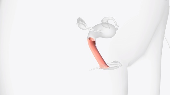
Free Images: "bestof:Gray1144.png The scrotum The penis has been turned upward and the anterior wall of the scrotum has been removed On the right side the spermatic cord the"
ST_PAULINUS_CHURCH.jpg
CHURCH_HOUSE_YALDING_KENT_ENGLAND.jpg
Gray1144.png
Esquelet humà frontal.svg
Design_Analysis_of_the_P-38_Lightning.png
Frans_Hals_-_Portrait_of_Jacob_Olycan_-_Mauritshuis_459.jpg
Aerial_photo_looking_W_at_Memorial_Amphitheater_under_construction_-_Arlington_National_Cemetery_-_Arlington_County_VA_USA_-_1919.jpg
Design_Analysis_of_the_Lockheed_PV-1_Ventura.png
Design_Analysis_of_the_Boeing_B-17G_Flying_Fortress.png
Gray1143.png
Layers_of_the_scrotum.gif
Design_Analysis_of_the_Vought_F4U-1D_Corsair.jpg
Gray1013.png
Gray1013_az.png
Gray516.png
Human placenta uterine side.jpg
Human placenta both sides.jpg
PZSL1907Page0895.png
Italian_-_Crucifix_-_Walters_71421.jpg
Hartington auditorium basketball court SW door.JPG
View of handyman drip art wall painting closeup on section.JPG
Workshop_of_Jacopo_del_Casentino_-_The_Last_Judgment_and_the_Crucifixion_-_Walters_37722.jpg
Human placenta baby side.jpg
Hartington auditorium upper lobby face N.JPG
GeorgeHerbertUprightVersionEasterWings1633.jpg
FMIB_47583_Solea_vulgaris_-_General_view_of_the_musculature_of_the_common_sole_seen_from_the_right_side,_after_removal_of_the_skin_The.jpeg
Egyptian_-_Wall_Fragment_of_Men_with_Bows_-_Walters_2298.jpg
The_Quarterly_journal_of_the_Geological_Society_of_London_(12736385365).jpg
FMIB_52753_Anatomy_of_the_internal_organs_of_a_female_gastropod,_viewed_from_the_dorsal_side_The_roof_of_the_mantle_cavity_has_been_divided.jpeg
Crucifixion-vita-mors-uta-codex-clm-13601-f3v-c1025.jpg
Wooden Alley.JPG
Recent memoirs on the Cetacea pl. 2.jpg
1920_Philadelphia_and_Columbia.jpg
Harbour_of_Christiania_(JW_Edy_plate_50).jpg
Christiania_great_Church_(JW_Edy_plate_51).jpg
Posidonius crater AS15-91-12366.jpg
Gastrointestinal Stromal Tumor (GIST) of Stomach.jpg
Bush book.jpg
Freeman's_Journal_27_January_1863_Letter_by_Timothy_Murphy.png
Terms of Use   Search of the Day