Click Here for More Images from iStock- 15% off with coupon 15FREEIMAGES 
Primary clear cell carcinoma of the pancreas is rare and it is classified as a \
Kidney cancer: Clear cell carcinoma. fuhrman nuclear grade-II, Renal growth shows a malignant tumor, renal cell carcinoma in between renal tubules.
Bronchitis and alveolitis in lung of horse infected with equine influenza virus, 200x magnification.
Natural agate surfaces, suitable for backgrounds and wallpapers.
Magnified sample of lung tissue displaying adenocarcinoma (cancer)
\
PAPS Smear study of a young women under microscopy showing Atrophic changes in uterus which is unusual at young age.
Quantum Gravity 3d rendered image of a comprehensive exploration of Quantum Gravity theories. String theory, loop quantum gravity (LOG), causal dynamical triangulation, twistor theory, gravitational waves, spacetime, gravity waves, quantum mechanics, quantum entanglement concepts.
Mucosa of a human stomach showing: surface epithelium, which invaginates to form the gastric pits. The fundic glands open into the pits and have parietal (pink) and chief (blue) cells.
Focal nodular hyperplasia (FNH) is a benign liver lesion that is composed of a proliferation of hyperplastic hepatocytes surrounding a central stellate scar. Typically, FNH is a solitary lesion that is more commonly seen in women.
Histopathology of diffuse sclerosing glomerulonephritis, light micrograph, photo under microscope
Slice of natural crystal for patterns and textures use with high resolution.
Breast tissue adenocarcinoma immunohistochemistry. Estrogen receptors.
Hyaline cartilage, Elastic cartilage and Bone Human under the microscope in Lab.
\
Thyroid cancer: Microscopic image of Metastatic papillary carcinoma of thyroid. Reactive change. Lymph node carcinoma.
Serous carcinoma is the most common type of ovarian cancer, accounting for approximately 75% of epithelial ovarian cancers.
Human lymph node. Metastasis of a malignant melanoma. The normal lymphoid tissue of lymph node has completely disappeared and has been replaced by malignant melanoma pigmented and non-pigmented cells.
Close-up shooting of a green agate stone mineral surface isolated on white background. Its a macro shot shows nature beautiful details with back lit. Agate is a kind of natural gemstone. Abstract pattern. Good for Wallpaper texture background. Purple
Bladder Cancer: High grade transitional cell carcinoma (TCC), Urothelial carcinoma
Acute ordinary hepatitis liver biopsy under light microscopy
Backgrounds of Bacteria and Mold under the microscope in Lab.
Microscopic image of Microscopic image of nymphaea of aqustio stem
Kidney cancer: Microscopic image of metastatic clear cell carcinoma of kidney, the most common type of renal cell carcinoma. Show brain tissue of malignant neoplasm, large atypical bizarre cells.
Mesenchymal dysplasia. Site: Placenta
Glomerulus affected by a severe capillary congestion. The glomerular capillaries are full of red blood cells. Human kidney. H&E stain\
Histological section of kidney tissue taken with microscope. Nuclei stain blue and cell cytoplasm is pink. Scattered red blood cells. Mostly tubules with two glomeruli present. Hematoxylin and Eosin (H&E) stain. More tissue sections:
Photomicrograph of Invasive squamous cell carcinoma of the nipple, Grade-II
Germ cell tumor of testicle under light microscopy zoom in different ranges
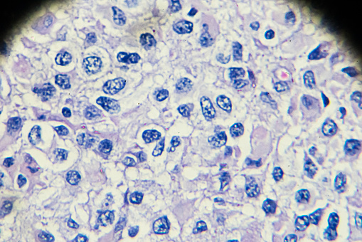
Free Images: "bestof:Granular Cell Tumor.jpg Granular Cell Tumor This 2-cm tumor presented as an abdominal wall mass thought to be an incisional hernia in a middle-aged woman Hard"
PET-MIPS-anim.gif
WHAT_YOUR_KIDS_ARE_BEING_TAUGHT.jpg
Pieter_Brueghel_the_Elder_-_The_Dutch_Proverbs_-_Google_Art_Project.jpg
Bi-specific T-cell engager.svg
Notable mutations.svg
THE_TRUTH_ABOUT_HILLARY.jpg
portugues-sexo_alemão-sex_ingles-sex_africano-seks_russo-секс_albanes-sex_chines-性別_bulgaro-секс_croata-spol_gales-rhyw_coreano-섹스_frances-sexe_japones-セックス_filandes-sukupuolesta_arabe-الجنس_prontos_ai_tens_em_15_linguas_a_p.jpg
Ascribed_to_Michelangelo_-_Christ_and_the_woman_of_Samaria_-_Google_Art_Project.jpg
Journal_of_Emerging_Infectious_Diseases_Jan_2013.jpg
Granular Cell Tumor.jpg
Krukenberg Tumor.jpg
Malignant lymphoma, high grade B cell 1.jpg
Gastrointestinal Stromal Tumor (GIST) of Stomach.jpg
Mature cystic teratoma of ovary 2.jpg
Hamartoma of the spleen.jpg
Warthin's tumor.jpg
Pheochromocytoma.jpg
Oncocytoma of the Salivary Gland.jpg
BCARS; Mouse Tissue (14803866837).jpg
Serous Cystadenocarcinoma of the Ovary.jpg
Endometriosis, abdominal wall.jpg
Cervical Carcinoma with Adnexa.jpg
Hhv-6.jpg
Nanoparticles; Medicine (5941040746).jpg
Hhv-6 (1).jpg
Müllerian adenosarcoma of the uterus.jpg
Hepatocellular carcinoma 1.jpg
Metastatic Melanoma in Lymph Node.jpg
Image_from_page_553_of_"The_diagnosis,_pathology_and_treatment_of_diseases_of_women_including_the_diagnosis_of_pregnancy"_(1868).jpg
Image_from_page_554_of_"The_diagnosis,_pathology_and_treatment_of_diseases_of_women_including_the_diagnosis_of_pregnancy"_(1868).jpg
Giant cell tumor of bone08.JPG
Mixed Tumor of the Salivary Gland.jpg
Aszites pleurexsudat.jpg
K055Rj_Dr._Alexander_Kemp_and_a_recovered_patient.jpg
Large_cell_carcinoma_of_the_lung.jpg
Pulmonal_multinodular_metastasis_of_a_renal_cell_carcinoma.jpg
Small_cell_carcinoma_of_the_lung.jpg
Well_differentiated_squamous_cell_carcinoma.jpg
Clear_cell_adenocarcinoma_of_the_lung.jpg
Squamous_cell_carcinoma_involving_a_subsegmental_bronchus.jpg
B-cell_chronic_lymphocytic_leukemia_(B-CLL).jpg
Faggot_cell_in_AML-M3.jpg
Large_cell_carcinoma_of_the_lung_.jpg
Computer with microarray.jpg
Microarray.jpg
Two_hypogranular_neutrophils_and_a_red_blood_cell_precursor_with_multiple_nuclear_fragments_in_RAEB.jpg
TNFB+TNFR 1TNR.png
Life_in_Motion_Fig_44.png
radiationstemcells_091109_01_sz_hr2400x1600.jpg.jpg
Macrophages in the brain before and after in vitro infecti.jpg
HHV-6_inclusion_bodies.jpg
Adenocarcinoma of the stomach.jpg
Rosette.jpg
Multiple Carcinoid Tumors of the Small Bowel 1.jpg
PTRS168_0715.png
Image_from_page_177_of_"Postoperative_treatment;_an_epitome_of_the_general_management_of_postoperative_care_and_treatment_of_surgical_cases_as_practised_by_prominent_American_and_European_surgeons"_(1907).jpg
Wilms Tumor CTScan.gif
Clara_Jacobi-Tumor.jpg
Ovarian surface papillary serous tumor submerged in water.jpg
FMIB_39125_Brook_trout,_showing_tumor_mass_at_the_tip_of_the_lower_jaw.jpeg
Woman_in_cell,_playing_solitaire.jpg
Pilomatricoma.jpg
Hepatocellular carcinoma 2.jpg
Adrenal_cortical_carcinoma.JPG
120627-O-ZZ999-004 (7467708342).jpg
K074R_Quessua_Hospital_-_Patient_with_a_large_lipoma.jpg
Abdominal Xray with uretal stent.jpg
Follicular adenoma of the thyroid.jpg
Infiltrating ductal carcinoma of the breast.jpg
Illu_blood_cell_lineage.jpg
US_Navy_060901-N-9865S-043_Dental_Officer_Lt._Cmdr._Chris_Pace_of_Waterloo,_Ill.,_performs_a_jaw_tumor_biopsy_in_the_ship^rsquo,s_medical_department_aboard_the_nuclear_powered_aircraft_carrier_USS_Nimitz_(CVN_68).jpg
Illu_blood_cell_lineage_(pt).png
Breast Cancer; Nanotubes; Antibodies (5880985350).jpg
CT scans (5941057880).jpg
PET-MIPS anim frame.PNG
Cajal_Retina.jpg
Villous adenoma of the sigmoid colon 1.jpg
Biopsy.jpg
Patients,_survivors_and_family_members_participate_in_Oncology_on_Canvas_130419-N-PJ759-004.jpg
Patients,_survivors_and_family_members_participate_in_Oncology_on_Canvas_130419-N-PJ759-003.jpg
Multiple Carcinoid Tumors of the Small Bowel 3.jpg
Patients,_survivors_and_family_members_participate_in_Oncology_on_Canvas_130419-N-PJ759-001.jpg
Multiple Carcinoid Tumors of the Small Bowel 2.jpg
Adenocarcinoma of the stomach 2.jpg
Digital Readout (8622935270).jpg
Intravascular Leiomyomatosis of the Uterus.jpg
AFIP405740R-SUBEPENDYMOMA.jpg
US_Navy_090804-N-9564W-035_Yeoman_1st_Class_Timothy_Gilbert,_assigned_to_Headquarters_Company_of_Naval_Mobile_Construction_Battalion_(NMCB_74),_donates_blood_during_a_blood_drive.jpg
Anthrax toxins (10444900713).jpg
The_Quarterly_journal_of_the_Geological_Society_of_London_(13859696235).jpg
Human Lung Cells (6680207077).jpg
Gray3.png
Wilhelm_Keitel_at_Nuremberg_Trials_November_1945.jpeg
Joachim_von_Ribbentrop_in_his_cell_Nuremberg_Trials_1945.jpeg
Nazi_party_member_Alfred_Rosenberg_in_cell_Nuremberg_Trials.jpeg
Maison_D'Arrét_Cellulaire_-_Galerie_Cellulaire.jpg
Bartolomeo Bellano - Saint Jerome in the Wilderness - Walters 54241.jpg
Nuremberg Trials defendant Wilhelm Frick in his cell 1945.jpeg.jpg
Terms of Use   Search of the Day