MAKE A MEME
View Large Image
| View Original: | Malignant lymphoma, high grade B cell 1.jpg (550x465) | |||
| Download: | Original | Medium | Small | Thumb |
| Courtesy of: | commons.wikimedia.org | More Like This | ||
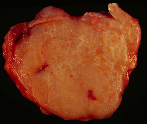
| Keywords: Malignant lymphoma, high grade B cell 1.jpg Non-Hodgkin's Lymphoma This elderly woman had carried a diagnosis of low-grade lymphoma since 1989 She had responded well to therapy and had been free of clinical evidence of lymphoma until spring 2000 when she presented with this inguinal mass Because she had developed a second cancer breast in the interim the question arose as to which if either cancer had recurred The clinicians opted for an excisional biopsy of the mass The specimen was 6 cm in greatest diameter soft and fleshy A quick touch prep showed that it was a lymphoma and fresh tissue was sent for surface marker characterization by flow cytometry The tumor cells which were gated in the medium and large areas of the cytogram marked as B cells with light chain restriction The cytologic features indicated a high-grade proliferation with a high mitotic rate although there was some debate as to whether it was a large- or small-cell follicular cell lymphoma it surprises those outside the field that such a basic question as cell size can be so controversial Ultimately my diagnosis was malignant lymphoma high grade B cell not otherwise specified To illustrate the difference in color quality between different types of lighting I shot the same frame under tungsten light this picture and window light see Image Malignant lymphoma high grade B cell 2 jpg Photograph by Ed Uthman MD Public domain Posted 19 May 00 http //web2 airmail net/uthman/specimens/index html PD Ed Uthman Gross pathology of neoplasms Diffuse large B cell lymphoma | ||||