Click Here for More Images from iStock- 15% off with coupon 15FREEIMAGES 
Fasciola hepatica, or liver fluke, 3D illustration. A parasitic trematode worm that causes fasciolosis, an infection of liver
plant Zea Stem C.S. under light mircoscope with white background
Striped mosquitoes life circle with white background. Life cycle of mosquitos isolate on white background.
Photomicrograph of filamentous blue-green algae, Oscillatoria species. Each band along the stand is one cell. This filament is very short probably having recently parted from a longer one. Filaments are typically many times longer. This algae is mobile, it moves and twists through the water. San Francisco Bay, California, USA. Live specimen. Wet mount, 40X objective, transmitted brightfield illumination. Note - motion blur of live specimen, very shallow depth of field, chromatic aberration and uneven focus are inherent in light microscopy.
Freshwater Closterium algae (unicellular charophyte green algae) - optical microscope x200 magnification
Molecular Model of Human Insulin with disulfide bonds shown.
Spirogyra sp. green algae under microscopic view - Chlorophyta, Green algae
3d plant Cell structure
Hand fungal infection. Ringworm, tinea manuum and close-up view of dermatophyte fungi Microsporum canis, Microsporum audouinii, Trichophyton rubrum and Trichophyton mentagrophytes, 3D illustration
Bacteria pathogen - 3d rendered microbiology image. Illustrate of Lactobacillus Bulgaricus Bacteria, Microbacterium Tuberculosis Bacteria (MTB). Medical research, health-care concept. SEM (TEM) scanning view
the tail of Aelurostrongylus abstrusus, cat lungworm under the microscope
3D rendering of microscopic particles and bubbles
Egg of intestinal fluke in human stool, analyze by microscope, original magnification 400x
DNA and biotechnology concept
Halofantrine is a drug used to treat malaria. Halofantrine's structure contains a substituted phenanthrene, and is related to the antimalarial drugs quinine and lumefantrine. 3d illustration
The protein responsible for Alzheimer's disease has recently been identified. It has been suggested that plaques formed by amyloid-β sheets may interfere with transmission at neuronal synapses. This image uses images of the plaques and the atrophied brain caused by Alzheimer's disease as a backdrop to illustrate the horror of the disease and the realistic phenomenon in vivo.
Colonies of bacteria grown from human skin smear, Staphylococcus epidermidis, left, and Micrococcus luteus, right, photo and 3D illustration
Close-up shot of a petri dish for checking clear zone an antibiotic in the laboratory.
Licmophora sp. algae, marine and freshwater diatom under microscopic view. Genus of benthic, photosynthetic and epiphyte diatom
Monkeypox Virus. 3D Render
centropyxis aculeata (amoeboid organism) under the microscope - optical microscope x400 magnification
Chain or ribbon diatom, a type of algae.
Squalane is a hydrocarbon derived by hydrogenation of squalene. 3d illustration
Structure of DNA in a Petri dish
Probiotics for organism. Microbiome in immune system. Human healthcare background. 3d-rendering
Molecular Model of Vitamin E.
Microalgae under microscopic view, green algae, cyanobacteria, phytoplankton, diatom, algae mix collage background
A ball and stick model of Eicosapentaenoic Acid or EPA. It is an omega-3 polyunsaturated fatty acid. It is introduced into the diet by eating oily fish. Isolated on white.
Folic acid, folate molecule. It is known as vitamin B9. Molecular model. 3D rendering. Illustration
Medical, Heredity, Chromosome DNA molecule double helix spiral genetic health concept in 3D illustration: Genetic mutation and genetic biotechnology disorders. Medical science, chemistry biology, gene cell polygonal wire-frame DNA background with copy space.
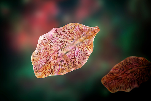
Free Images: "bestof:Fasciola LifeCycle(French version).gif en Causal Agents The trematodes Fasciola hepatica the sheep liver fluke and Fasciola gigantica parasites of herbivores"
BlankMap-World6.svg
Fasciola_LifeCycle(French_version).gif
Fasciola_LifeCycle.gif
Fasciola_LifeCycle_2013.png
Fasciola hepatica (Linnaeus, 1758) 2013 000-2.jpg
Opisthorchis_LifeCycle.gif
Babesia_LifeCycle(French_version).png
Fasciola hepatica (Linnaeus, 1758) 2013 001-2.jpg
Fasciolopsis_buski_LifeCycle.gif
Opisthorchis_LifeCycle(French_version).GIF
Malaria_LifeCycle(French_version).GIF
Life-cycle-of-fasciola-gigantica.png
Fasciola hepatica.jpg
Egg_of_Fasciola_hepatica_08G0041_lores.jpg
Fasciola_hepatica_cycle_in_Czech.jpg
Fasciola hepatica2.jpg
Idiacanthus_fasciola1.jpg
Clonorchis_sinensis_LifeCycle.png
Toxocara_canis_LifeCycle(French_version).GIF
Terms of Use   Search of the Day