Click Here for More Images from iStock- 15% off with coupon 15FREEIMAGES 
Bone marrow plasma cells .Plasma cell neoplasms are diseases in which the body makes too many plasma cells. Plasma cell neoplasms can be benign or malignant. Diagnosed with Immunostain CD138 positive.
High Vaginal Swab (HVS) Gram Stain: Photomicrograph showing Bacterial vaginosis (BV).
Biochemical analysis in the laboratory
Characteristics of Rhizopus is a genus of common saprophytic fungi  on Slide under the microscope for education.
Thyroid cancer: Microscopic image of Metastatic papillary carcinoma of thyroid. Reactive change. Lymph node carcinoma.
Photomicrograph of filamentous blue-green algae, Oscillatoria species. Each band along the stand is one cell. This filament is very short probably having recently parted from a longer one. Filaments are typically many times longer. This algae is mobile, it moves and twists through the water. San Francisco Bay, California, USA. Live specimen. Wet mount, 40X objective, transmitted brightfield illumination. Note - motion blur of live specimen, very shallow depth of field, chromatic aberration and uneven focus are inherent in light microscopy.
Close-up shot of a petri dish for checking an antibiotic in the laboratory.
Lily anther c.s. under microscope
Close up shot of a gloved hand touching a centrifuge tube holding solutions with a pH meter probe in it.
Photomicrograph of Histological analysis of Intramedullary SOL showing Astrocytoma, WHO grade-4
Microbiology lab work in a research facility. Hands of a lab technician spreading bacterial culture over the nutritional agar
Diffuse large B-cell lymphoma, or DLBCL, is a cancer that starts in white blood cells called lymphocytes.
Cross sections of the plant root under the microscope view for education
plant Leaf of Nerium indicum T.S. under light mircoscope with white background
Breast tissue adenocarcinoma immunohistochemistry. Estrogen receptors.
stx2a is a subtype of Shiga toxin-producing E. coli (STEC) linked to Hemolytic Uremic Syndrome (HUS), a severe condition impacting kidney function and blood clotting. STEC poses a significant risk of foodborne illness and can spread within communities including family members.
Patient lying on the examination table receiving his dose of collagen and vitamins in injection applied directly to the face
PAPS Smear study of a young women under microscopy showing Atrophic changes in uterus which is unusual at young age.
The study of plant tissues under the microscope in the laboratory.
Photomicrograph of two rotifers. Rapidly rotating cilia at top of heads, internal organs visible. Live specimen. Wet mount, 10X objective, transmitted brightfield illumination.
Microscopic image of ankle joint fluid. Osteochondroma disease. Osteochondrama is the most common benign tumor of bone. show bony chips and cartilage.
A conceptual image of an abstract network of multi-coloured organic strands arranged in a non regular pattern all connected together in a complex manner. The network is made from smaller multi-coloured squares and polyhedra, in vivid colours against a pastel toned background. Shows background and foreground elements to the structure.
Light micrograph of the anterior surface of a heavily pigmented human iris lined by stromal fibroblasts and abundant melanocytes. It is one of the few body surfaces not lined by an epithelium. Beneath is the iris stroma also with many melanocytes. This iris belongs to a deep brown or black eye.
Neuroendocrine carcinoma well differentiated (carcinoid) rectum with immunostain synaptophysin
Liver Fluke Section under light microscope with white background
Photomicrograph of Histological analysis of Intramedullary SOL showing Astrocytoma, WHO grade-4
Cross-section Dicot, Monocot and Root of Plant Stem under the microscope for classroom education.
Close-up shot of a petri dish for checking an antibiotic in the laboratory.
Stratified flat epithelium under microscope
Digital Dispersed corona virus on petri dish. Animation of multiplication of CORONA VIRUS COVID-19
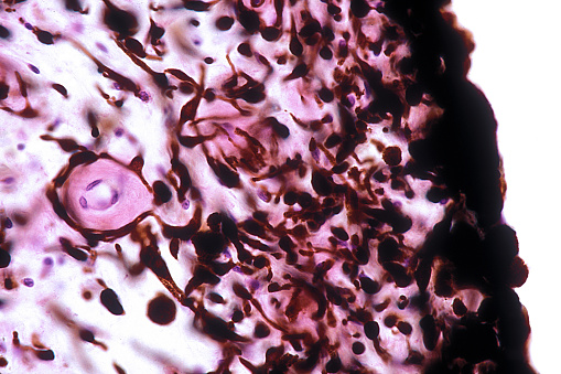
Free Images: "bestof:Bone marrow biopsy.jpg W Biopsie cs Kostní dřeň kostní dřeně cs A from that page 021204-N-0696M-180 W Georgetown University Hospital Washington D C Dec 4"
surgery-operation-hospital-patient-85574.jpg
Pieter_Brueghel_the_Elder_-_The_Dutch_Proverbs_-_Google_Art_Project.jpg
Second_Congregational_Church_NRHP_71000020.jpg
Notable mutations.svg
Olvany 4101089538 9c4b2723e3 o.jpg
Pittsburgh newspaper consolidation timeline.svg
Bone_marrow_biopsy.jpg
US_Navy_021204-N-0696M-165_Surgeon_Hans_Janovich_performs_a_bone_marrow_harvest_operation_on_Aviation_Electronics_Technician_1st_Class_Michael_Griffioen.jpg
US_Navy_021204-N-0696M-171_Surgical_technician_Amina_Sherali_places_recently_transfused_bone_marrow_from_Aviation_Electronics_Technician_1st_Class_Michael_Griffioen_into_a_sterile_bag_in_preparation_for_transplant.jpg
FMIB_47350_Chub_or_Fall_Fish,_Semotilus_bullaris,_Bone_Pond,_Franklin_Co,_N_Y_Dec_5,_1911.jpeg
Georgetownuniversityhospital.JPG
Hospital Bohunice.jpg
US_Navy_071212-N-2735T-059_Hospital_Corpsman_2nd_Class_Antoinette_Peterson_takes_a_swab_sample_from_Aviation_Machinist's_Mate_3rd_Class_Roger_Cunningham_to_submit_for_a_bone_marrow_drive_aboard_the_amphibious_assault_ship_USS_N.jpg
Waseda knihovna.jpg
Bohuslav_Stoces_1.jpg
Tomas_Trnka_1929.jpg
Julius_Komárek_(1892-1955).jpg
KTF UK.jpg
Populus_alba5.jpg
Illiac II Control Panel.jpg
AF8E4701 (8122031384).jpg
AF8E4667 (8122009435).jpg
AF8E4698 (8122011383).jpg
AF8E4680 (8122029178).jpg
AF8E4672 (8122010333).jpg
AF8E4695 (8122025332).jpg
AF8E4685 (8122005781).jpg
Zcu fel.jpg
Georges_Cuvier_by_George_Thomas_Doo.jpg
7.díl html m3a8ddb32.png
Thomas_Hogan.jpg
1855_Colton_Plan_or_Map_of_Washington_D.C._-_Geographicus_-_WashingtonDC2-colton-1855.jpg
Alois_Velich.jpg
FMIB_48926_Plaice.jpeg
2007 US News Top 40 colleges.png
The_Tusoulan_villa_Loudon.jpg
FMIB_46272_Mysis_relicta,_one_of_the_Mysidacea.jpeg
FMIB_34362_Bone_Coming_on_Board.jpeg
Css_hunley_on_pier.jpg
Frantisek_Tilser.jpg
FMIB_44687_Scrimshawed_Whale's_Teeth_Engraved_by_Whalemen.jpeg
James_R._Browning.jpg
Charlotte_Salomon_-_JHM_4762_-Kristallnacht.jpg
The_Quarterly_journal_of_the_Geological_Society_of_London_(12981002405).jpg
FMIB_44662_Right_Whaling_Cutting-In_the_Bone.jpeg
FMIB_44688_Jagging_Wheels,_carved_from_Whale's_Teeth,_Bone,_Etc,_by_the_Whalemen.jpeg
FMIB_34361_Bone_on_Deck_and_Blubber_Coming_on_Board.jpeg
FMIB_49778_Bone_wedding_ring,_made_as_though_the_bride_and_groom_both_wore_it_during_the_ceremony.jpeg
Toronto_Magnetic_Observatory_in_1852_by_William_Armstrong.jpg
Ford_A4206_NLGRF_photo_contact_sheet_(1975-04-27)(Gerald_Ford_Library).jpg
Ford_A4208_NLGRF_photo_contact_sheet_(1975-04-27)(Gerald_Ford_Library).jpg
Charlotte_Salomon_-_JHM_4175.jpg
Charlotte_Salomon_-_JHM_4175T.jpg
FMIB_38661_Col_William_C_Gorgas;_the_Hospital_Grounds,_Ancon.jpeg
Charlotte_Salomon_-_JHM_4175TC.jpg
Charlotte_Salomon_-_JHM_4155.jpg
FMIB_44455_Glimpses_of_the_Exposition_Grounds_and_Views_of_the_Various_Buildings_Completed_in_the_Fall_of_1908_Picturestaken_Eight_Months.jpeg
FMIB_41331_Fort_Shoemaker,_Dall_River_Log_House_Intended_for_Use_as_a_Hospital_but_its_Occupancy_was_Never_Found_to_be_Necessary.jpeg
Wartburg-Laute 001.jpg
Classification of images progress human.png
Everything_for_the_garden_(Page_55)_(16647110765).jpg
Image_from_page_281_of_"Canadian_grocer_July-December_1895"_(1895).jpg
Image_from_page_72_of_"Ontario_Sessional_Papers,_1877,_No.1-3"_(1877).jpg
Ford_A4207_NLGRF_photo_contact_sheet_(1975-04-27)(Gerald_Ford_Library).jpg
Belgique_horticole_(10329186155).jpg
United_States_Exploring_Expedition_(Pl_19)_(7052041087).jpg
Terms of Use   Search of the Day