Click Here for More Images from iStock- 15% off with coupon 15FREEIMAGES 
Human anatomy detail of shoulder. Muscle, bone structure, arteries. On plain studio background. Professional lighting.
Defined back muscles of Asian female
The trapezius muscles, commonly known as the traps, are a pair of large, triangular-shaped muscles located in the upper back and neck region. They are among the most prominent muscles of the upper body and play a crucial role in various movements involving the shoulders and neck.
3d spine model on a uniform gray background
The thoracic spine is the second segment of the vertebral column, located between the cervical and lumbar vertebrae .
Digital medical illustration: Human spine featuring vertebrae (cervical (C1-C7), thoracic (T1-T12) and lumbar (L1-L5)) vertebrae, discs and pelvis.
3D Rendering of Male Full Body Deep lntrinsic Back Network Muscles Rear View Isolated on White Background with Skeletal Bones and Text Labeling
A digital radiograph is used to produce a high-precision analog X-ray film. It is used for diagnostics in the field of internal and external diseases.
An acupuncture model of the male human torso depicting the acupoints and meridiens used in Traditional Chinese Medicine and massage therapy
3D illustration, neck painful - cervical spine skeleton x-ray, medical concept.
The chest muscles, specifically the pectoralis major and pectoralis minor, are important muscles located in the anterior (front) part of the chest. They play a significant role in various upper body movements, particularly those involving the shoulder and arm.
X-Ray Image Of Human Spine for a medical diagnosis. The red part indicates the center of pain.
Muscles of the Back Anatomy isolated on white background. 3D render
MRI scanning of the human body skeletal, muscular and nervous systems with abdomen region view. / You can see the animation movie of this image from my iStock video portfolio. Video number:
Labeled human anatomy Erector Spinae back muscles in isolation on the skeletal system from a posterior view on a white background.  3D rendering.
waist pain three Dimensional
Young woman stretching over isolated white background. Image taken with Hasselblad H3D camera system and developed from camera RAW.
The costovertebral joints describe two groups of synovial plane joints which connect the proximal end of the ribs with their corresponding vertebrae
Human Muscle Anatomy For medical concept 3D Illustration
Computer futuristic digital HUD software interface with a system for scanning the human body and transmitting images and signals in real time. Within one complex, both a scanner and a digital video recorder are used.
The gastrocnemius muscles, commonly referred to as the calf muscles, are a pair of powerful muscles located in the back of the lower leg. They are the most superficial muscles of the calf and play a crucial role in various movements involving the ankle and knee joints.
Studio shot of an unrecognizable young sportswoman stretching against a grey background
Man's upper body muscles frontal anterior view labeled on a white background.
Xray view of a patient with an injured spine and suffering. / You can see the animation movie of this image from my iStock video portfolio. Video number: 1453180480
Woman with back pain, vertical
The posterior sacroiliac ligament is a compound ligament composed of three distinct bands 3d illustration
3D Illustration Concept of Human Muscular System Torso Muscles Trapezius Muscle Anatomy
Abstract staircase detail seen from below, forming a spiral with golden proportions. White walls with copper orange elements resembling steps, almost giving feeling of vertigo. Abstract architectural background with copy space.
Human back muscles isolated
white textile covered mannequin for measuring and applying the dress while sewing and cutting processes by a tailor
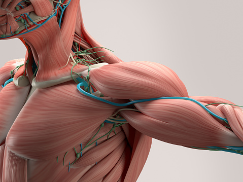
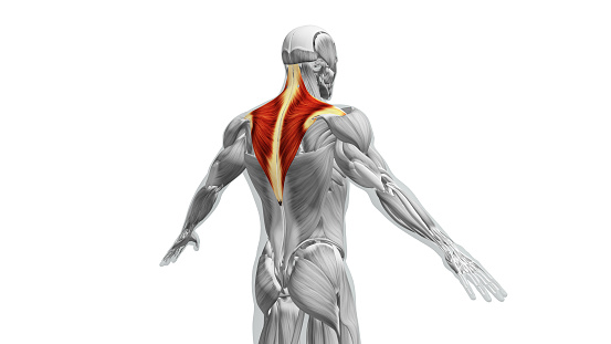
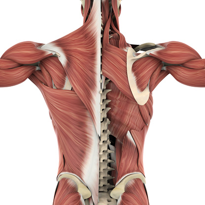
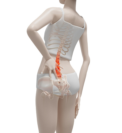
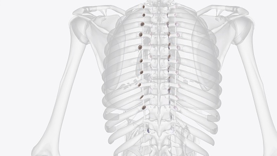
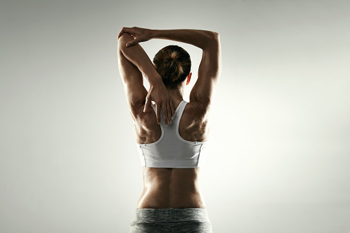
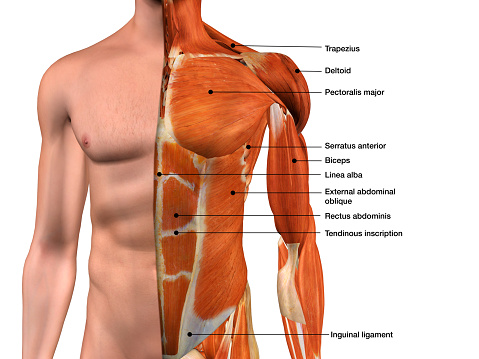
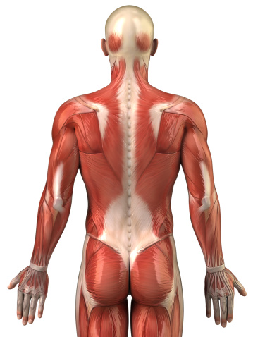
Free Images: "bestof:Back Muscles.jpg Posterior view of muscles connecting the upper extremity to the vertebral column A Trapezius B Teres Major C Teres Minor D Latissimus Dorsi E"
Shoulder joint.svg
Shoulder_joint-es.svg
Shoulder_joint-pt.svg
Design_Analysis_of_the_Lockheed_PV-1_Ventura.png
Gray814_pl.svg
Dermatomes and cutaneous nerves - posterior.svg
Sobo_1909_260.png
Gray512.svg
George_Stubbs_-_A_Comparative_Anatomical_Exposition_of_the_Structure_of_the_Human_Body_with_That_of_a_Tiger_and_a_Co..._-_Google_Art_Project_(2345936).jpg
Design_Analysis_of_the_P-38_Lightning.png
Back_Muscles.jpg
Teres_major.PNG
Latissimus_dorsi.PNG
Supraspinatus.PNG
Nuchal_ligament.PNG
Lumbar_triangle.PNG
Armens_muskler_2,_Nordisk_familjebok.png
Trapezius_Gray409.PNG
Serratus_posterior.PNG
Deltoideus_posterior.PNG
Scm_posterior.PNG
Musculus_rhomboideus_minor.PNG
Musculus_rhomboideus_major.PNG
External_oblique_posterior.PNG
Gray409serratusposteriorinferior.png
Gray412.png
Gray523.png
Gray203.png
Gray207.png
Gray809.png
Gray409.png
Infraspinatus.PNG
Iliac_crest.PNG
Superior_nuchal_line.PNG
Musculus_splenius_capitis_et_cervicis.png
Musculus_splenius_capitis_et_cervicis_marked.png
Gray525.png
Arm_muscles_back_numbers.png
Gray1211.png
Gray208.png
Gray576.png
Gray818.png
Gray129.png
Gray810.png
Gray521.png
Armens_muskler_1,_Nordisk_familjebok.png
Teres-major.png
Teres-minor.png
Gray832.png
Grays_Anatomy_image392.png
Gray235.png
Gray808.png
Gray235-ar.png
PZSL1907Page334.png
PZSL1907Page333.png
PZSL1907Page336.png
Shoulderjoint.PNG
Gray800.png
Nerves_of_the_left_upper_extremity.gif
Gray827.png
PZSL1889Page012.png
The_Quarterly_journal_of_the_Geological_Society_of_London_(12736562693).jpg
The_Quarterly_journal_of_the_Geological_Society_of_London_(12960130963).jpg
ChronicBackache1037.png
ChronicBackache1042.png
The_muscles_of_the_left_leg,_seen_from_the_front,_and_the_bones_and_muscles_of_the_right_leg_seen_in_right_profile,_and_between_them,_a_patella._Drawing_by_Michelangelo_Buonarroti,_ca._1515-1520._WDL3249.png
Illu_upper_extremity_muscles.jpg
Gray950_az.png
Gray950_az_Qalxan.png
Gray950_az_Üzük.png
Gray959_az.png
Gray960_az.png
Gray950_az_Qirtlaq_qapagi.png
Gray950_az_Çalov.png
Gray813.png
Gray1215.png
Gray814.png
Rectus_capitis_posterior_major.png
Rectus_capitis_posterior_minor.png
Gray812.png
Braus_1921_85.png
FMIB_47895_Left_crusher_claw_of_lobster,_partly_dissected_from_upper_side,_to_show_relations_of_muscles,_nerves,_blood_vessels,_and_skin.jpeg
Gray165.png
Gray410.png
Dermatomes and cutaneous nerves - posterior.png
PZSL1907Page0887.png
PZSL1907Page0888.png
FMIB_47896_Fig1--Leftsecond_periopod_from_anterior_or_upper_side,_partly_dissected_to_show_the_relations_of_muscles_and_tendons_in_the.jpeg
Gray1231.png
Gray1232.png
Gray960_muscles_of_larynx_ru.png
Leonardo_da_Vinci_-_Anatomical_studies_of_the_shoulder_-_WGA12824.jpg
Armens_muskler_3,_Nordisk_familjebok.png
Pelorosaurus_conybeari.jpg
Gray700.png
Gray1036.png
Horse_anatomy_posterior_view.jpg
ChronicBackache1038.png
Cetiosaurus_brevis.jpg
Dacentrurus_dorsal_vertebra.jpg
Terms of Use   Search of the Day