Click Here for More Images from iStock- 15% off with coupon 15FREEIMAGES 
After dopamine binding, the dopamine receptor changes his shape and binds the inactive G protein.The G protein expels its GDP molecule replacing it by GTP. and falls into two pieces. The Galpha subunit binds to and activates the enzyme adenylyl cyclase, which produces then cyclic AMP spreading a signal through the cell.
Chemical formula of Progesterone
3D cartoon and Gaussian surface models, chain id color scheme, PDB 2y73, white background
Molecular Model of Vitamin D.
Copper(II) sulfate with molecular structure. Chemical ingredient used in medical and public health issues.
A molecular model of the flavonoid Quercetin.  An antioxidant and anti-inflamatory, it is used as a nutritional suplement.  It is commonly found in many fruits and vegetables and is a significant chemical component of tea.  Isolated on white.
Folic acid, folate molecule. It is known as vitamin B9. Molecular model. 3D rendering. Illustration
Colorful geometric shapes connected with arrows forming a software diagram.
Ferrocene is an organometallic compound. The molecule consists of two cyclopentadienyl rings bound on opposite sides of a central iron atom. 3d illustration
Chemical formula of Cortisol
3D cartoon and Gaussian surface model, chain id color scheme, PDB 3v7t, white background
3D realistically coloured hexagonal shapes of connected spheres, background with copy space. Futuristic illustration design template for scientific presentation. Molecular business, science, teamwork concept.
A GTP-bound G-alpha protein (blue) binds to and activates the enzyme adenylyl cyclase, which produces then cyclic AMP spreading a signal through the cell.
Molecular model of lactose, a disaccharide sugar in milk.
Hyaluronan, hyaluronic acid, HA, hyaluronate molecule, short fragment. Molecular model. 3D rendering. Illustration
A model of a molecule of lysine, an amino acid. Amino acids are the building blocks of proteins and have many functions in metabolism.
Cediranib cancer drug molecule. 3D rendering. Atoms are represented as spheres with conventional color coding.
Chemical formula of Aldosterone
3D cartoon and Gaussian surface models, chain id color scheme,  PDB 2iw2, white background
Rendered coronavirus/2019-nCoV single-stranded RNA, built from experimentally determined Ebola virus crystal structure. Color symbols - bluewhite=carbon, pink=oxygen, lightblue=nitrogen, white=hydrogen, lavender=phosphorus
The glucagon like peptide-1 receptor (GLPr) is a G proteincoupled receptor (GPCR) that mediates the action of GLP-1, a peptide hormone. The activated receptor  has a strong effect  on the management of type 2 diabetes mellitus and obesity, including glucose homeostasis and regulation of gastric motility and food intake.
Molecular model of Adenosine triphosphate,  or Adenosine-5'-triphosphate (ATP)
Remdesivir is a broad-spectrum antiviral medication, it is a specific treatment for COVID-19. 3d illustration
3D realistically coloured hexagonal shapes of connected spheres, background with copy space. Futuristic illustration design template for scientific presentation. Molecular business, science, teamwork concept.
Filtration and water purification scheme contaminated molecules pass through filter layers
Liquid bubbles up Fluid Twirl
3D cartoon and Gaussian surface models, PDB 6vg8, white background
A model of a molecule of Glycine, an amino acid. Amino acids are the building blocks of proteins and have many functions in metabolism.
Vitamin A. Retinol 3d molecule on paper texture background. Skeletal formula of vitamin a. Recessed text in green and yellow.
Conceptual background with colourful spheres positioned in tension.
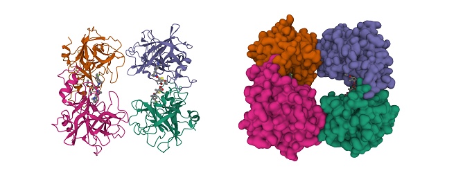
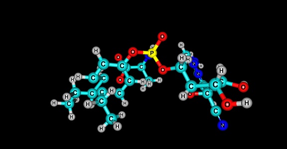
Free Images: "bestof:ACAC mechanism.png The reaction mechanism of ACAC A B The color scheme is as follows enzyme coenzymes substrate names metal ions phosphate and carbonate"
ACAC mechanism (zh-cn).svg
Nucleotides syn1.svg
P450cycle.svg
Simple mechanism.svg
Transamination mechanism (zh-cn).svg
Reaction scheme competitive enzyme inhibition.svg
Reaction scheme enzyme inhibition 1.svg
Reaction scheme enzyme inhibition 2.svg
Reaction scheme uncompetitive enzyme inhibition.svg
Reaction scheme non-competitive enzyme inhibition.svg
Bromoacetamidation mechanism.svg
Acetyl-CoA-Carboxylase.svg
Seleniumdioxide oxidation.svg
Selenoxide elimination.svg
Skraup quinoline synthesis mechanism.svg
Benzoin condensation scheme.svg
Simple mechanism - PT.svg
Vicarious nucleophilic substitution.svg
Cysteinprotease Reaktionsmechanismus.svg
Epoxidation Mimoun Mechanism.svg
Design_Analysis_of_the_Boeing_B-17G_Flying_Fortress.png
Design_Analysis_of_the_B-24J_Liberator.png
Design_Analysis_of_the_F6F-5_Hellcat.png
ACAC mechanism.png
Kynureninase reaction.png
Simple mechanism of enzyme reaction (zh-tw).png
Simple mechanism of enzyme reaction zh.png
Aldolase b reaction mechanism.jpg
Chemical mechanism of phosphoglucose mutase.png
Allan-Robinson reaction mechanism.png
Michaelis-Arbusov reaction mechanism.png
Woodward cis-Hydroxylation Reaction Mechanism.png
DFMO mechanism.png
Scheme of the E2 reaction mechanism.JPG
Rubottom Oxidation Mechanism.png
Aromatase mechanism.png
Homoserine Dehydrogenase Hydride Transfer Reaction Mechanism.jpg
Mechanism for the binding of ATP and then Ubiquitin substrate to the ubiquitin activating enzyme.png
BVR mechanism.png
Scheme of the E2 elimination reaction mechanism - the second step.JPG
Scheme of the E2 elimination reaction mechanism.jpg
Scheme of the E1 elimination reaction mechanism - the first step.JPG
Streptokinase mechanism of action.png
CBDO Reaction Scheme.png
NOS reaction.png
RNR reaction.png
Barton-Zard mechanism.png
Succinyl CoA Synthetase Mechanism Revised.png
Mecanismo de reacción de Barton Kellogg.png
Mecanismo de la síntesis de Biginelli.png
Vinylcyclopropane rearrangement concerted.png
Mecanisme simple.png
Nucleophilic Epoxidation Mechanism2.tiff
H2OAutoIonization.png
SN2-MeSH-MeI-2D-A.png
Proposedmechanismforbglii.gif
SN2-MeSH-MeI-2D-B.png
Huygens_clock.png
ChronicBackache1034.png
Design_Analysis_of_the_Vought_F4U-1D_Corsair.jpg
Terms of Use   Search of the Day