Click Here for More Images from iStock- 15% off with coupon 15FREEIMAGES 
\
Photomicrograph of Histological analysis of Intramedullary SOL showing Astrocytoma, WHO grade-4
Dust mites,Dermatophagoides pteronyssinus
View of a flea under a microscope.
Close up view of a cat flea seen under a microscope
Dog Fleas on a Flea Comb after combing them off of a dog, against a white background
Microscopic fungi Malassezia furfur, showing yeast cells and hyphae. dermatophytes, Nail scraping or skin scraping for fungus test in microbiology laboratory.
Cheyletiella blakei Mite - Small Cat pest view under a microscope
pest control treatment, german cockroach trapped on a sticky paper sheet
Rare Scorpion (Arachnida) inclusion in Burmese amber, Myanmar, Cenomian, Upper Cretaceous, approximately 100 million years ago, age of dinosaurs. Image taken with extreme macro and focus stacking technique.
Dog flea under microscope. 40x magnification. Ctenocephalides canis
plant Zea Stem C.S. under light mircoscope with white background
Flea cat's parasite close-up macro
Compound eyes of the fly and hairy body, extreme close-up
ctenocephalides felis under the microscope, cat flea
Hashimoto thyroiditis is an autoimmune disorder involving chronic inflammation of the thyroid.
3D rendering of animal fleas
Close up of small feeding tick with shiny pinhead. Nikon D7200 with Nikon 105mm macro lens & extension tube, SB 21 flash. Photo taken in Gainesville, Florida
Malt Extract Agar in Petri dish using for growth media to isolate and cultivate yeasts, molds and fungal testing from Neil scraping samples. Neil scraping for fungus culture.
A macro image of a house fly.
Olive Fruit fly- Bactrocera oleae: one of the most important olive pests and it parasitoid (ichneumonid wasp).
Photomicrograph of two rotifers. Rapidly rotating cilia at top of heads, internal organs visible. Live specimen. Wet mount, 10X objective, transmitted brightfield illumination.
A Corixidae nymph under the microscope and in polarized light.
Portrait of Blow Fly on blue
House Fly Leg W.M. under light mircoscope with white background
centropyxis aculeata (amoeboid organism) under the microscope - optical microscope x400 magnification
Photograph of a prepared flea slide.
Side View Of A Fly Isolated On A White Background
Algae cells under microscope magnification 10X
Simuliidae, black fly (Diptera) in Baltic amber, Eocene, approximately 56 - 34 million years ago. Image taken with extreme macro and focus stacking technique.
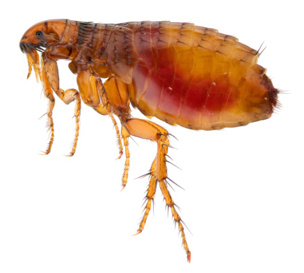
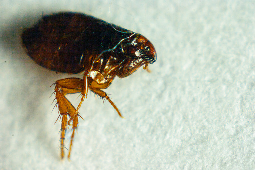
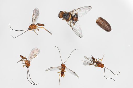
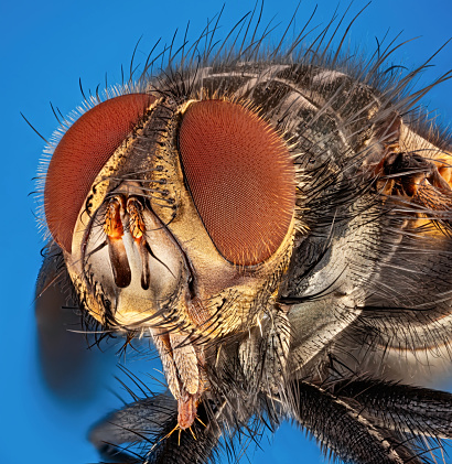
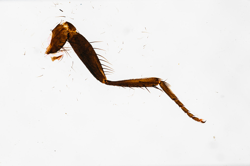
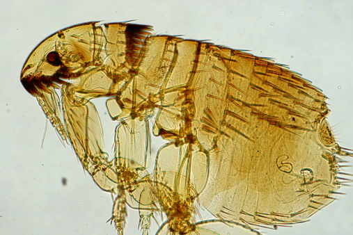
Free Images: "bestof:flea siphonaptera insect parasite electron microscopy electron micrograph magnification false color"
flea-siphonaptera-insect-parasite-63043.jpg
mite-tuckerella-acari-arachnid-67582.jpg
epidermis-leaf-electron-microscopy-63195.jpg
bacteria-salmonella-pathogens-67659.jpg
bacteria-cocci-staphylococci-67791.jpg
crystals-ice-crystal-snow-ice-67792.jpg
ice-crystal-crystals-snow-ice-67709.jpg
snow-crystals-ice-ice-crystal-67708.jpg
bacteria-disease-escherichia-coli-62993.jpg
koli-bacteria-escherichia-coli-123081.jpg
mite-aceria-anthocoptes-acari-67638.jpg
Ebola_Virus_Particles.jpg
Laughlin quasiparticle interferometer sample scanning electron micrograph.svg
Multistable current controlled negative resistance.svg
Surface ionization of cesium.svg
Scanning_Electron_Micrograph_of_a_Flea.jpg
Nanoparticles; scanning electron micrograph (5887833046).jpg
Electron_Micrograph_of_Flea_derivate.jpg
Schistosoma 20041-300.jpg
Layered Approach May Yield Stronger, More Successful Bone Implants (5880495301).jpg
Microscopy, Scanning Photoionization Imagery, SPIM Image (5940488089).jpg
Blood clot in scanning electron microscopy.jpg
Baculovirus.jpg
Hemozoin crystals.jpg
Yersinia_pestis.jpg
Yersinia_pestis_Bacteria.jpg
Insect SEM gracilariidae.jpg
H5N1_virions_108,000x.jpg
Nanoknife.jpg
Micrococcus_luteus_9758.jpeg
Hhv-6 (1).jpg
Anthrax_color_enhanced_micrograph.JPG
HeLa-IV.jpg
MRSA_dead_neutrophil.jpg
Malaria_Plasmodium_gallinaceum.jpg
Conductive probe SEM images.png
Silicon Rings (10747924673).jpg
Nanoimprint Lithography Tests (5940484963).jpg
Nanoimprint Lithography Tests (5887832648).jpg
Velvet_ant_stinger_(tip)--scanning_electron_microscope_image.jpg
HIV_H9_T-cell_II.jpg
Titanium Dioxide TiO2 (8051508739).jpg
A_(H5N1)_virion,_a_type_of_bird_flu_virus_which_is_a_subtype_of_avian_influenza_A.jpg
Melanoma_sem.jpg
Velvet_ant_head_anterior_--_SEM_photo.jpg
Red_blood_cell_infected_with_malaria_parasites-NIAID_RML.jpg
Nanoparticles (6283421358).jpg
TEM; Silver Nanoparticles (5978792602).jpg
White_blood_cell_with_MRSA.jpg
3-D Nanostructures (5887271107).jpg
TEM of silicon nanowire (5887830328).jpg
Schistosome_Parasite_SEM.jpg
Drosophilidae compound eye.jpg
Nanowires that Emit UV Light (5884303101).jpg
Drosophilidae compound eye edit1.jpg
Surface_of_HIV_infected_microphage.jpg
Dendritic_cell_revealed.jpg
SEM_Lymphocyte.jpg
Transmission line animation.gif
Electron asymmetric motion animation.gif
Solar AM0 spectrum with visible spectrum background (en).png
Solar AM0 spectrum with visible spectrum background (no).png
Terms of Use   Search of the Day