Click Here for More Images from iStock- 15% off with coupon 15FREEIMAGES 
Blue colored stem cells of a lentil plant under the microscope.
Carcinoid tumors are a type of slow-growing cancer that can arise in several places throughout your body. Carcinoid tumors, which are one subset of tumors called neuroendocrine tumors, usually begin in the digestive tract (stomach, appendix, small intestine, colon, rectum) or in the lungs. Site: Ovary
Psoriasis, dermatologic diseases.
Quantum Gravity 3d rendered image of a comprehensive exploration of Quantum Gravity theories. String theory, loop quantum gravity (LOG), causal dynamical triangulation, twistor theory, gravitational waves, spacetime, gravity waves, quantum mechanics, quantum entanglement concepts.
Histological Pancreas human, Liver human, Vermiform appendix human and Kidney Human under the microscope for education.
PAPS Smear study of a young women under microscopy showing Atrophic changes in uterus which is unusual at young age.
3d rendering of Fluid painting, abstract colorful liquid texture background.
Abstract chaotic oil painting. Texture and background artistic.
Squamous cell carcinoma of the lung is a type of lung cancer. It occurs when abnormal lung cells multiply out of control and form a tumor.
Microscope photo of a sambucus stem with parenchyma cells.
plant Pine Cone-Female L.S. under light mircoscope with white background
Cross sections of the plant root under the microscope view for education
Abstract background Organic Form - 3d rendered image of glass structure, optical technology, nano electrical wave, dispersion colorful rainbow.  Organic shapes and form.  Science fiction or organic technology concept.
Prostate Cancer: Microscopic image of prostatic tissue, adenocarcinoma, malignant neoplasm, atypical epithelial cells.
Close up of texture and colors of the bottom of a burnt pot.
Stratified flat epithelium under microscope
Plasmacytoid urothelial carcinoma (PUC) is a unique variant of invasive urothelial carcinoma characterized by tumor cells that exhibit a striking resemblance to plasma cells, and it is known to have an aggressive behavior.
Host cells with spores (mold) are inside wood under the microscope for education.
Closeup abstract color mixing of acrylic for use as background. Acrylic texture with marble pattern, blue marbling background
Light micrograph of an unstained iris showing the localization of pigment cells. The melanin pigment is concentrated in the back surface of the iris stroma, near the pigment epithelium (right). Between both, there is a clear band that corresponds to the iris dilator muscle. The anterior surface shows a lower concentration of pigment.
Gastric mucosa intestinal metaplasia under microscopy
Photomicrograph of Histological analysis of Intramedullary SOL showing Astrocytoma, WHO grade-4
This is a color photograph detailing different body parts of a caucasian man in his early 40s with wet skin after being in a hot tub.
Human male beard hair close-up view
Lymphocytic myocarditis. Myocarditis is an inflammation of the heart muscle. Lymphocytic refers to the type of cell seen in the affected tissue. A lymphocyte is a type of immune function cell.
Reticular cell is a type of fibroblast. They are found in many tissues including the spleen, lymph nodes and lymph nodules. Pathology of human.
Abstract marble texture background stock photo, suitable for design.
Education anatomy and Histological sample of Human under the microscope.
Yellow abstract background
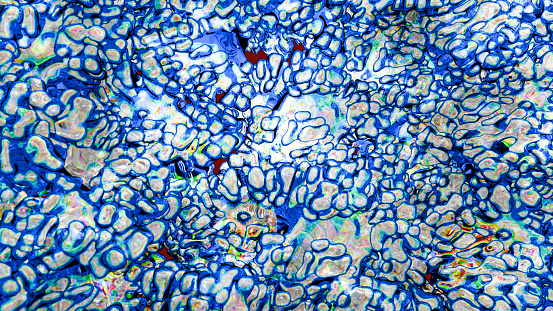
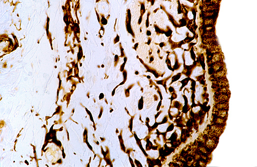
Free Images: "bestof:Wangiella dermatitidis PAS stain PHIL 3781 lores.jpg This micrograph reveals the histopathologic changes in phaeohyphomycosis due to Wangiella dermatitidis"
Wangiella_dermatitidis_PAS_stain_PHIL_3781_lores.jpg
Conidiophore_of_the_fungus_Bipolaris_hawaiiensis_PHIL_4338_lores.jpg
Histopathologic_features_of_aspergillosis_including_the_presence_of_conidial_heads_PHIL_4335_lores.jpg
Schistosoma_haematobium_egg_4842_lores.jpg
Taenia_solium_tapeworm_proglottids_5261_lores.jpg
Adult_Trichuris_female_human_whipworm_PHIL_414_lores.jpg
Terminal_chlamydospores_of_the_fungus_Microsporum_audouinii_PHIL_3054_lores.jpg
Gravid_proglottid_from_the_cestode_Taenia_saginata_5259_lores.jpg
Head_of_Enterobius_vermicularis_human_pinworm_5230_lores.jpg
Schistosoma_mansoni_egg_4841_lores.jpg
Terms of Use   Search of the Day