Click Here for More Images from iStock- 15% off with coupon 15FREEIMAGES 
Prostate Cancer: Microscopic image of prostatic tissue, adenocarcinoma, malignant neoplasm, atypical epithelial cells.
Quantum Gravity 3d rendered image of a comprehensive exploration of Quantum Gravity theories. String theory, loop quantum gravity (LOG), causal dynamical triangulation, twistor theory, gravitational waves, spacetime, gravity waves, quantum mechanics, quantum entanglement concepts.
Tissue of Small intestine (Duodenum), Large intestine Human and Stomach Human under the microscope in Lab.
Chronic appendicitis pathology under light microscopyzoom in different areas.
Metastatic plasmacytoma, metastatic undifferentiated sarcoma. Microscopically show cellular material of mostly plasmacytiod cells. atypical mitoses are present. plasma cell tumor.
A microscope slide showing a stained section of a human bowel, or small intestine.  It was taken through microscope lens that has a central focus through a thin layer of glass.
Carcinoid tumors are a type of slow-growing cancer that can arise in several places throughout your body. Carcinoid tumors, which are one subset of tumors called neuroendocrine tumors, usually begin in the digestive tract (stomach, appendix, small intestine, colon, rectum) or in the lungs. Site: Ovary
Leiden leaf c.s. under microscope
PAPS Smear study of a young women under microscopy showing Atrophic changes in uterus which is unusual at young age.
Microscopic image of Microscopic image of nymphaea of aqustio stem
The study of plant tissues under the microscope in the laboratory.
Digital Dispersed corona virus on petri dish. Animation of multiplication of CORONA VIRUS COVID-19
linden stem (Tilia platyphyllos) cross section under the microscope - optical microscope x200 magnification
Acute ordinary hepatitis liver biopsy under light microscopy
traditional thai fabric
Lymphocytic myocarditis. Myocarditis is an inflammation of the heart muscle. Lymphocytic refers to the type of cell seen in the affected tissue. A lymphocyte is a type of immune function cell.
Cross sections of the plant root under the microscope view for education
corn stem micrograph with dye
Host cells with spores (mold) are inside wood under the microscope for education.
3d rendering of Fluid painting, abstract colorful liquid texture background.
Photomicrograph of Histological analysis of Intramedullary SOL showing Astrocytoma, WHO grade-4
Tilia Stem C.S.under light microscopy
Plants cell texture.
Metastatic neuroendocrine tumor. Site: Liver.  Neuroendocrine tumors are cancers that begin in specialized cells called neuroendocrine cells. Neuroendocrine cells have traits similar to those of nerve cells and hormone-producing cells. Neuroendocrine tumors are rare and can occur anywhere in the body.
Background, background image,blue polka dot background picture
Hyaline cartilage, Elastic cartilage and Bone Human under the microscope in Lab.
\
Illustration of coloured texture forming a background
Closeup abstract color mixing of acrylic for use as background. Acrylic texture with marble pattern, blue marbling background
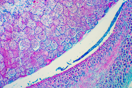
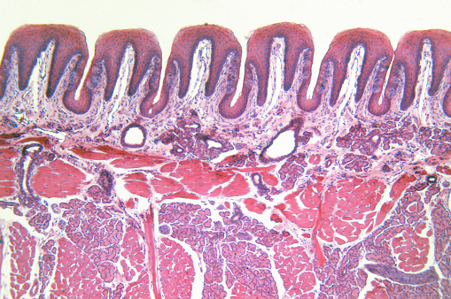
Free Images: "bestof:Villous adenoma of the colorectum (high power view).jpg en Microscopically the diagnosis of a villous adenoma is easily confirmed by observation of the enlarged"
Villous adenoma of the colorectum (high power view).jpg
Villous adenoma of the colorectum (medium power view).jpg
Villous adenoma of the colorectum (low power view).jpg
Tubulovillous Polyp of the Colon 1.jpg
Villous adenoma of the sigmoid colon 1.jpg
Villous adenoma of the sigmoid colon 2.jpg
Villous adenoma of the sigmoid colon 3.jpg
Villous adenoma of the sigmoid colon, gross pathology.jpg
Terms of Use   Search of the Day