Click Here for More Images from iStock- 15% off with coupon 15FREEIMAGES 
forming of underskin cellulite illustration
A Computed Axial Tomography scan of my brain.  Taken with contrast.  Great image for medical applications.
CAT Scan image pulmonary angiography showing lung adenocarcinoma with red arrow. Medical themes
Computational tomography image of the abdominal and pelvic regions.
PAPS Smear study of a young women under microscopy showing Atrophic changes in uterus which is unusual at young age.
Mammogram
Diffuse large B-cell lymphoma, or DLBCL, is a cancer that starts in white blood cells called lymphocytes.
Dicot plants stem show plant vascular tissue under light microscope view for botany education.
MRI of the prostate gland, revealing an enlarged size, aids in diagnosing tumors, guiding treatment decisions, and monitoring prostate health.
Human brain MRI.
Generic blue neuron cell model isolated on a white background with copy space. Science, neuroscience, biology, microbiology, neurology 3d rendering illustration.
Quantum Gravity 3d rendered image of a comprehensive exploration of Quantum Gravity theories. String theory, loop quantum gravity (LOG), causal dynamical triangulation, twistor theory, gravitational waves, spacetime, gravity waves, quantum mechanics, quantum entanglement concepts.
Medical Imaging and Radiology
abstract alloy texture design background.
CT Scan of an abdomen of a patient with abdominal pain showing swollen appendix suggesting the diagnosis of acute appendicitis.
MRI scan of veins around the heart
CTA Coronary artery  3D rendering image. CT angiography for heart Vessel disease .
Serous carcinoma is the most common type of ovarian cancer, accounting for approximately 75% of epithelial ovarian cancers.
Cat Scan of female patient with centrilobular emphysema. Medical themes
CT of thorax of four patients having covid-19 disease. Four variations are seen on four lung x-ray images. Shot with a full frame mirrorless camera.
The lymph nodes are small, bean-shaped organs located throughout the body .
CAT scan of human abdomen...man with aneurysm of abdominal aorta
anatomy of the posterior view of heart with attached great vessels
Computational tomography image of the abdominal and pelvic regions.
CTPA or CTA pulmonary artery .This imaging technique offers a clear view of the pulmonary arteries, aiding in the diagnosis of pulmonary embolism, vascular conditions, and other respiratory issues.
Carcinoid tumors are a type of slow-growing cancer that can arise in several places throughout your body. Carcinoid tumors, which are one subset of tumors called neuroendocrine tumors, usually begin in the digestive tract (stomach, appendix, small intestine, colon, rectum) or in the lungs. Site: Ovary
X-ray multiple part of human and many medical condition and disease .
Section of epididymis under light microscope with white background
Eye disease, eye cancer, 3d illustration
Close-up shooting of a green agate stone mineral surface isolated on white background. Its a macro shot shows nature beautiful details with back lit. Agate is a kind of natural gemstone. Abstract pattern. Good for Wallpaper texture background. Purple
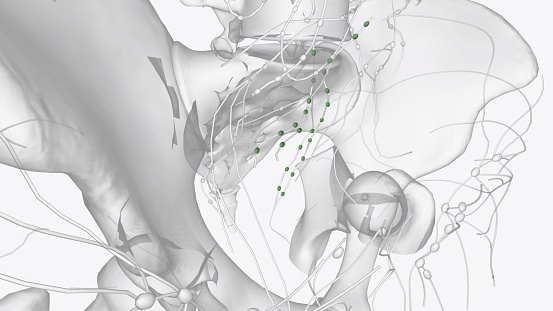
Free Images: "bestof:Variation 4 of internal iliac artery branching.svg Internal iliac artery branching variations - 2008-01-23 Made for the sake of free knowledge to all mankind by"
Terms of Use   Search of the Day