Click Here for More Images from iStock- 15% off with coupon 15FREEIMAGES 
Squamous cell carcinoma or squamous cell cancer (SCC or SqCC) is a cancer of a kind of epithelial cell, the squamous cell. These cells are the main part of the epidermis of the skin, and this cancer is one of the major forms of skin cancer. However, squamous cells also occur in the lining of the digestive tract, lungs, and other areas of the body, and SCC occurs as a form of cancer in diverse tissues, including the lips, mouth, esophagus, urinary bladder, prostate, lung, vagina, and cervix, among others. Despite sharing the name squamous cell carcinoma, the SCCs of different body sites can show tremendous differences in their presenting symptoms, natural history, prognosis, and response to treatment. Micrograph of squamous cell carcinoma of the head and neck
Photomicrograph of Histological analysis of Intramedullary SOL showing Astrocytoma, WHO grade-4
Showing Light micrograph Type of Tissue Human under the microscope in Lab.
Magnetic resonance images of the brain (MRI brain). Computed resonance imaging of the human brain.
Cat Scan of female patient with centrilobular emphysema. Medical themes
\
Thyroid cancer: Microscopic image of Metastatic papillary carcinoma of thyroid. Reactive change. Lymph node carcinoma.
Atmospheric photo of modern MRI scanning room in hospital. Non-invasive examination. Magnetic resonance imaging technology in specialized medical clinic.
Virus and bacteria infected the Human lungs, lung disease, 3d illustration
Serous carcinoma is the most common type of ovarian cancer, accounting for approximately 75% of epithelial ovarian cancers.
A student in a medical institution is studying the topic of lungs. Pandemic. Home schooling. Pulmonary disease. World Asthma and Allergy Day. World Tuberculosis Day
Mammogram
Photomicrograph of Histological analysis of Intramedullary SOL showing Astrocytoma, WHO grade-4
CAT Scan lung lobe
Adipose tissue human, Smooth muscles human, Bone human and Striated (skeletal) muscle human under the microscope in Lab.
Computational tomography image of the abdominal and pelvic regions.
Astrocytoma, Intramedullary SOL (C5-D3) tissue biopsy. Photomicrograph show malignant neoplasm of oval to elongated cells with eccentric cuclei and eosinophilic cytology. Micovascular proliferation.
Carcinoid tumors are a type of slow-growing cancer that can arise in several places throughout your body. Carcinoid tumors, which are one subset of tumors called neuroendocrine tumors, usually begin in the digestive tract (stomach, appendix, small intestine, colon, rectum) or in the lungs. Site: Ovary
MRI of the prostate gland reveals Focal abnormal SI lesion at left PZpl at apex as described; PI-RADS category 4, clinically\nsignificant cancer is likely.
PAPS Smear study of a young women under microscopy showing Atrophic changes in uterus which is unusual at young age.
Kidney proximal tubule epithelial cloudy SWELLING under different area in light microscopyKidney proximal tubule epithelial cloudy SWELLING under different area in light microscopyKidney proximal tubule epithelial cloudy SWELLING under different area in light microscopy
Histological Pancreas human, Liver human, Vermiform appendix human and Kidney Human under the microscope for education.
Photo of throax cat scan image of Coronavirus (covid-19) positive patient is seen on computer monitor while a doctor is seen at the end of bright tunnel in radiology clinic. Radiologist is wearing a hazmat protective suit, protective gloves, face mask and eyewear and using a digital tablet. Shot indoor with a full frame mirrorless camera.
Metastatic plasmacytoma, metastatic undifferentiated sarcoma. Microscopically show cellular material of mostly plasmacytiod cells. atypical mitoses are present. plasma cell tumor.
Adult granulosa cell tumor. Site: ovary. Granulosa cell tumor of the ovary is a rare type of ovarian cancer. This type of tumor is known as a sex cord-stromal tumor and usually occurs in adults.
Human lung pathology under light microscope, The lungs is organs of the respiratory system in human. Human pathology education. Haematoxylin and eosin staining technique slide.
Histological section of kidney tissue taken with microscope. Nuclei stain blue and cell cytoplasm is pink. Scattered red blood cells. Mostly tubules with two glomeruli present. Hematoxylin and Eosin (H&E) stain. More tissue sections:
Photomicrograph of Stomach Adenocarcinoma. Gastric Adenocarcinoma.
A microscope slide showing a stained section of a human bowel, or small intestine.  It was taken through microscope lens that has a central focus through a thin layer of glass.
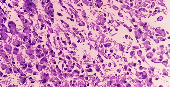
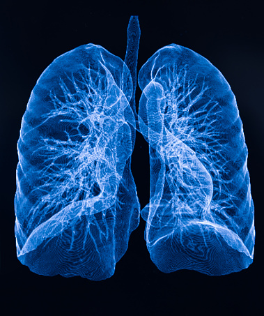
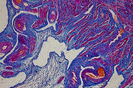
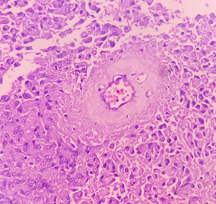
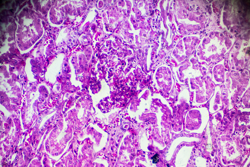
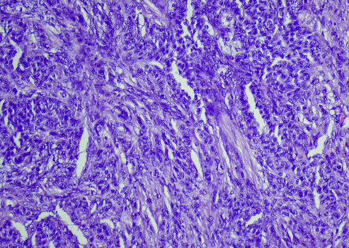
Free Images: "bestof:UIPlungbiopsy.jpg en Appearance of usual interstitial pneumonia UIP in a surgical lung biopsy at low magnification The tissue is stained with hematoxylin purple"
UIPlungbiopsy.jpg
Honeycomb change.jpg
Fibroblast focus.jpg
Image_from_page_554_of_"The_diagnosis,_pathology_and_treatment_of_diseases_of_women_including_the_diagnosis_of_pregnancy"_(1868).jpg
Trichinella_rat_BAM2.jpg
Evermicularis_worm4_HB.jpg
Cdc_cjd.jpg
Egranulosus cysts HB.jpg
Everything_for_the_garden_(16854204875).jpg
Wholesale_price_list_(16189008607).jpg
Wholesale_price_list_of_requisites_for_florists_and_market_gardeners_(16775431562).jpg
Everything_for_the_garden_(16854182975).jpg
Terms of Use   Search of the Day