Click Here for More Images from iStock- 15% off with coupon 15FREEIMAGES 
fossil of a trilobite. lived during time of Early Cambrian throughout the lower Paleozoic, during the Devonian til the end of the Permian See also my other Fossils and Mineral/ Crystal images:
close up on Trilobite fossil isolated on white background
The upper pole of the left kidney lies at the level of the 12th thoracic vertebra, and the lower pole lies at the level of the third lumbar vertebra .
Haliotis kamtschatkana, northern abalone or pinto abalone, is a species of large sea snail, a marine gastropod mollusc in the family Haliotidae, the abalones. It has been listed as \
trilobite fossil texture as very nice natural background
Photomicrograph of planaria, a flatworm, Dugesia species. Slight motion blur in head. Freshwater stream, San Luis Obispo, California. Live specimen. Wet mount, 2.5X objective, transmitted brightfield illumination.
Microraptor gui fossil, Early Cretaceous.
Skeleton of a dead dog in a dry area
superior view of heart showing opening of aorta, superior vena cava, pulmonary trunk with related valve
Rudists embeded in sandstones, cross sections.\nCretaceous marine fossis near Barcelona.
Phacops rana (Eldredgeops rana) is a species of trilobite from the middle Devonian period
plant Pine Cone-Female L.S. under light mircoscope with white background
trilobite fossil texture as nice natural background
Fish skeleton on black background
Trilobite Paradoxides spiulosus
Knightia alta is an extinct genus of fish well-known from abundant fossils found in the Green River Formation of Wyoming, United States. They rarely exceeded 10 cm in length and are found throughout the formation. Knightia alta, Eocene Epoch, Green River Shale. Fossil.
four Fossils Specimens  : Practical Specimens for Study of Earth Science / Isolated white
Ammonite and Orthoceras Fossil preserved in the rocks mined from the reserve found in the Middle Atlas Mountains of Morocco
Thoracic Vertebral, T8 Twelve vertebrae are located in the thoracic spine and are numbered T-1 to T-12 3d illustration
Ammonite inside a limestone rock. The ammonites where living between 409 and 65 million years ago (Mya). The image was captured in the canton of Aargau (Switzerland).
Hand Drawn of Human Anatomy Cranial Bones on Chalkboard
Photomicrograph of segmented worm, Chaetogaster sp. Lives in ponds. Live specimen. Wet mount, 5X objective, transmitted brightfield illumination.
close up on Trilobite fossil isolated on white background
Ancient fish fossil in stone
Ancient fish fossils are on a rock
Ancient rock art incised in a sandstone boulder.
Trilobite fossil (Cheirurus ingricus) from the middle Ordovician peroid (488.3 – 443.7 million years ago) . This specimen was found in Russia
skin science 3d
Leiden leaf c.s. under microscope
Monotomidae, Coleoptera Fossil in Burmese amber of Cenomanian era, 100 million years ago, from the state of Myanmar, extreme macro shot
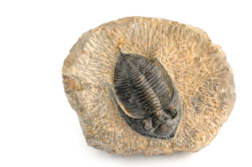
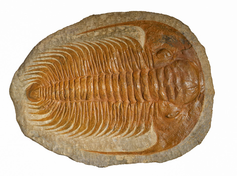
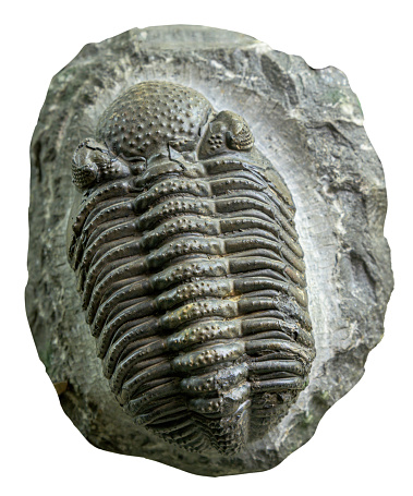
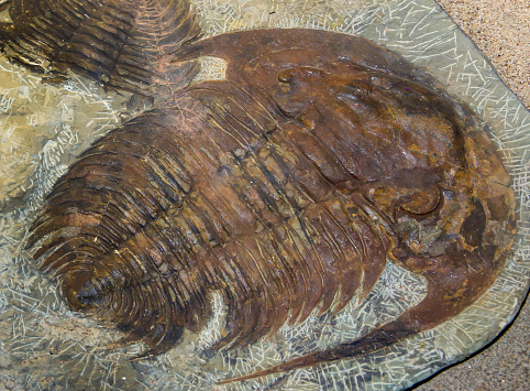
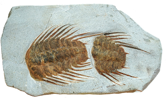
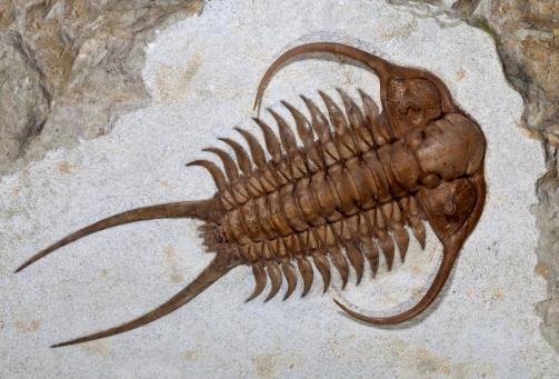
Free Images: "bestof:Trilobite cephalon ventral anatomy.png en Diagram showing the three major sections of a trilobite Cephalon Thorax Pygidium Trilobite_sections_numbered svg"
Trilobite sections numbered.svg
Trilobite sections-en.svg
Trilobite sections-ms.svg
Trilobites main morphological groups.svg
Trilobite cephalon areas-en.svg
Trilobite cephalon areas numbered.svg
Trilobite sections-ru.svg
Trilobite cranidium numbered.svg
Trilobite cranidium-en.svg
Trilobite lobes numbered.svg
Trilobite lobes-en.svg
Culex restuans larva diagram num.svg
Gray5.svg
Gray5_-_es.svg
Wignspan Diagram.svg
Incandescent light bulb.svg
Culex_pipiens_diagram_en.svg
6 week human embryo nervous system.svg
Culex pipiens diagram uk.svg
Ant worker morphology (ja).svg
Culex pipiens diagram fr.svg
Culex pipiens diagram es.svg
Gray5-ja.svg
German School system.svg
Culex pipiens diagram zh.svg
Linearly independent vectors in R3.svg
Gray5_de.svg
Gray5_nl.svg
Incandescent light bulb (no labels).svg
Project-triangle.svg
Human skull side suturas pl.svg
Human skull side details pl.svg
Human skull side details right pl.svg
Cross section jellyfish en.svg
Jellyfish cross section numbered.svg
Deutsches Schulsystem.svg
Gray769-pt.svg
FL barchart by number 2008-11-30.svg
Diagram of swine influenza symptoms EN.svg
Scheme ant worker anatomy-en.svg
Coax and Skin Depth.svg
Diagram of influenza infection symptoms EN.svg
Cambial Variants (Anomalous Secondary Growth) Fig. 8.5.1 Heteropteris.svg
Cambial Variants (Anomalous Secondary Growth) Fig. 8.5.3 Phaedranthus.svg
Cambial Variants (Anomalous Secondary Growth) Fig. 8.5.5 Ceropegia.svg
Cambial Variants (Anomalous Secondary Growth) Fig. 8.5.4 Passiflora.svg
Cambial Variants (Anomalous Secondary Growth) Fig. 8.5.2 Acacia pennata.svg
PD Diagram of swine flu symptoms EN.svg
Libyan Uprising-mk.svg
Design_Analysis_of_the_P-38_Lightning.png
Terms of Use   Search of the Day