Click Here for More Images from iStock- 15% off with coupon 15FREEIMAGES 
Virus isolated on white background
Oncolytic Virus immunotherapy and cancer vaccine Therapy as a treatment to kill cancers by attacking the malignant tumor cell and infecting them and destroying the pathogen as a 3D render.
Virus 3d model set. Covid-19 germs, fungi, bacteria objects. Graphic from microscopic zoom in lab for learning science medical, biology, virology on white background. Clipping path. 3D Illustration.
Coronavirus Covid-19
Human adenovirus model. Structure of the adenoviridae virion: penton base, nucleocapsid with DNA genome, capsomers, proteins. Acute respiratory viral infections, ARVI disease, 3D medical science mixed media illustration
Disease X Cells. 3D Render
A combined image of various micro organisms in colorhttp://195.154.178.81/DATA/istock_collage/0/shoots/785093.jpg
Adeno-associated viruses, 3D illustration. The smallest known viruses to infect humans, belong to the family Parvoviridae, are used for gene therapy, and for creation of isogenic human disease models
Haematococcus pluvialis red cyst algae under microscopic view, empty space - haematocyst, resting cells, strong antioxidant astaxanthin
COVID-19 new mutation variant - 3d rendered image. Pandemic wave.\nViral Infection concept. Delta, Omicron (B.1.1.529), Centaurus (B.1.1.529.2.75) (BA.2.75), Aeterna (BA.4.6) variant. Sars-cov-2, 2019-nCoV, Coronavirus.\nAntibody, Antigen, stealth, Vaccine technology concept.\nB117 - COVID-19 Variant,\nSARS-CoV-2 BA.2 Variant,\nSARS-CoV-2 BA.5 Variant,\nSARS-CoV-2 Omicron Variant,\nSaRS-CoV-2 Deltacron Variant
3D CG rendered image of scientifically accurate Adeno-Associated Virus (AAV) Capsid Structure based on PDB : 1LP3 (surface style)
A group of viruses together as seen under a microscope
3d illustration of Smallpox or Monkeypox viruses
Dengue virus cells. 3D Render
Nirmatrelvir inhibits the 3C-like protease of the SARS-CoV 2 virus. The coronavirus 3CL hydrolase enzyme, also known as the main protease, is essential for the maturation of the virus.
Virus cell isolated on the white background.  Scientific medical illustration.
3d illustration Influenza Virus or Virus
Particles, medical concept
Subvariant Virus Spreading as viral pathogens Mutating variants and mutation as a transmissible health risk concept and new COVID-19 outbreak or coronavirus mutations and influenza background as a 3D render.
Concept of COVID-19 cells, with blue background, and medicine pills around. Focus with depth of field. Cure concept render.
Dengue viruses, a group of RNA viruses transmitted by mosquitoes and responsible for causing dengue fever, 3D illustration.
Molecular model of amylose, one of the two sugar components in starch
3d rendering of The nanoemulsions (NE) are tiny molecules that carry the drug components in its core. The structure is organic surfactant, oil or active ingredient and co-emulsifier
Disease X Cells. 3D Render
Influenza virus diagram. 3D illustration.
The three-dimensional structure of proteins is represented in a variety of conformations. This image is based on the ribbon structure of a protein. It is suitable for biology notebooks and book covers.\nThis image has not been created by AI.
COVID-19 Corona virus with spike glycoprotein are floating on the air . Blue color background . 3D rendering .
COVID-19 new Omicron sub-variant BQ.1.1 - 3d rendered image. Pandemic wave.\nViral Infection concept. Delta, Omicron (B.1.1.529), Centaurus (B.1.1.529.2.75) (BA.2.75), Aeterna (BA.4.6) variant. Sars-cov-2, 2019-nCoV, Coronavirus.\nAntibody, Antigen, stealth, Vaccine technology concept.\nB117 - COVID-19 Variant,\nSARS-CoV-2 BA.2 Variant,\nSARS-CoV-2 BA.5 Variant,\nSARS-CoV-2 Omicron Variant,\nSaRS-CoV-2 Deltacron Variant
bacteria virus or germs microorganism cells. 3d rendering.
Natural agate surfaces, suitable for backgrounds and wallpapers.
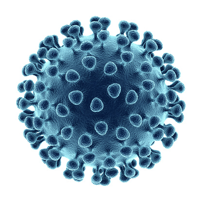
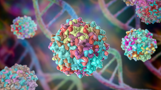
Free Images: "bestof:The structure of the immature HIV-1 capsid in intact virus particles.png en Structure of the Immature HIV-1 Capsid in Intact Virus Particles at 8 8A Resolution"
Ebola_Virus_Particles.jpg
Ebola_virus_particles_found_both_as_extracellular_particles_and_budding_particles_from_chronically-infected_African_green_monkey_kidney_cells_..jpg
bokeh-background-white-balls-red-973249.jpg
Hepatitis B virus v2.svg
7DMA.svg
PPV genome.svg
HIV-SIV-phylogenetic-tree1.svg
Particle chart Log.svg
Taxol structure.svg
dengue-virus-3d-structure-biology-1186407.png
dengue-virus-3d-structure-medicine-1186411.png
virus-bacteriophage-science-biology-154694.svg
hepatitis-b-virus-virus-3d-1186581.png
hepatitis-b-virus-virus-3d-1186583.png
hepatitis-b-virus-virus-3d-1186582.png
immune-system-blood-cells-virus-1132182.jpg
microbiology-dna-people-structure-163521.jpg
particles-wave-circle-color-1435366.jpg
particles-wave-circle-color-1435334.jpg
particles-wave-circle-color-1435354.jpg
particles-wave-circle-color-1435363.jpg
particles-wave-circle-color-1435356.jpg
particles-wave-circle-color-1435361.jpg
organic-render-fractal-menger-676367.jpg
t-helper-cell-cell-immune-system-1123292.png
particles-color-colorful-wave-1677652.jpg
Phenyldecahydroquinoline.svg
particles-abstract-glass-refraction-1038688.jpg
monitor-display-particles-color-1677621.jpg
Trimethylsilyl propionate.svg
tangle-fibers-about-hair-form-701993.jpg
star-particles-banner-header-1328394.jpg
star-particles-banner-header-1328393.jpg
star-particles-banner-header-1328392.jpg
particles-banner-header-background-1328400.jpg
monitor-display-particles-success-1677622.jpg
star-particles-heart-love-1340474.jpg
render-3d-bug-slug-bacteria-684129.jpg
Satratoxin-H.svg
cubes-mirror-reflection-cool-993101.jpg
binary-monitor-particles-blue-1414324.jpg
March_2,_1906.jpg
P450cycle.svg
Theodore_Gericault-_Evening_landscape_with_an_aqueduct,_1818.jpg
Endomembrane system diagram en (edit).svg
Tryptophan biosynthesis (en).svg
Animal mitochondrion diagram en.svg
Animal mitochondrion diagram en (edit).svg
The structure of the immature HIV-1 capsid in intact virus particles.png
Structure_of_HIV.jpg
Norwalk Caspid.jpg
Steven_H3N2_Flu_ET.jpg
Flu und legende color c.jpg
Gag Molecules (5888007689).jpg
Hiv-i.jpg
Azt.jpg
Azt (1).jpg
Papillomavirus capsid.png
H1N1_virus_particles_II.jpg
H1N1_virus_particles.jpg
Chikungunya_virus_particles-PHIL-17369.jpg
Nodamura Virus Structure.jpg
Sarmynd2.jpg
HI-Virion-gl.png
PDB 1m4x EBI.jpg
PDB 1w8x EBI.jpg
PDB 1dyl EBI.jpg
3EVA.png
P2-baseplate.tif
IPNV_structure.jpg
TMV.jpg
RSV secondary structure.jpg
Potato12zd.jpg
HIV_spike_in_complex.jpg
HI-virion.jpg
Ppv2.gif
Neuraminidase from 1918 influenza.gif
HIVRRE SecondaryStructure.png
US_Navy_040324-N-0130O-001_Hospital_Corpsman_1st_Class_Jason_Dillow,_of_Westminster,_Md.,_draws_blood_from_a_Sailor^rsquo,s_arm,_for_a_routine_human_immunodeficiency_virus_(HIV)_check.jpg
US_Navy_041118-N-8213G-016_Hospital_Corpsman_3rd_Class_Kasandra_Hopper,_from_Cumberland_Gap,_Tenn.,_verifies_the_name_on_the_blood_specimen_taken_for_human_immunodeficiency_virus_(HIV)_testing.jpg
HIV-1_protease_for_slides.png
Bourbon_virus_sphere_(EID_2015_Fig_2b).jpg
Jmol2WUX.pdb.jpg
Bubblegram.jpg
Bourbon_virus_endocytosis_(EID_2015_Fig_2d).jpg
HLA B^1501.png
CXCL12 1A15.png
Influenza M2 2RLF.png
Amyloid-beta-42 1IYT.png
Ta C structure.jpg
Spinal-disc-protrusion-l5.jpg
NS1 Dimer InfluenzaH5N1 3f5t bio r 500.jpg
Saint.Sepulcre.png
Eau_Claire_Town_Hall_and_Survey_Publishing_Company_Building_NRHP_79003371.jpg
CloudSat-CALIPSO_Launch_Successfully,_Image_of_the_Day_DVIDS749611.jpg
James_Monroe_Law_office_8a.gif
Syr-Darya_Oblast._City_of_Turkestan._Main_Niche_in_the_Mausoleum_Leading_to_the_Tomb_of_Saint_Sultan_Akhmed_Iassavi_WDL3586.png
Antiquities_of_Samarkand._Shrine_of_Chupan-Ata._General_View_from_the_Southwest_WDL3741.png
Antiquities_of_Samarkand._Mausoleum_of_Khodzha_Abdu-Derun._General_View_of_the_Mausoleum_from_the_Southwest_WDL3734.png
Terms of Use   Search of the Day