Click Here for More Images from iStock- 15% off with coupon 15FREEIMAGES 
Macro photo of human eye, iris, pupil, eye lashes, eye lids.
Cropped image of screen showing researcher looking in a microscope fertilising an egg. Intracytoplasmic sperm injection.
The study of plant tissues under the microscope in the laboratory.
Hokkigai on White Background
MRI scan of the  brain   for detect  Brain  diseases sush as stroke disease, Brain tumors and Infections.
Pink iris isolated on black background.
Anatomy of the sacrum bone, showcasing its intricate details and features, 3D illustration. Perfect for educational or medical purposes
Magnetic resonance images of the brain (MRI brain). Computed resonance imaging of the human brain.
Lady's Slipper orchid on black background
plant stem (dahlia stem) cross section under the microscope showing epidermis, bascular bundles (phloem and xylem) and cortex - optical microscope x400 magnification
Fresh raw clams on white background studio shot.
Isolated orchid flowers with clipping paths.
Microscopic image of the cross section of the root of a buttercup (crowfoot) plant - Ranunculus repens. The propeller shaped pattern in the center is the vascular tissue for transporting water and nutrients up and down the plant. The circles are the individual cells.
CTPA or CTA pulmonary artery .This imaging technique offers a clear view of the pulmonary arteries, aiding in the diagnosis of pulmonary embolism, vascular conditions, and other respiratory issues.
Leiden leaf c.s. under microscope
plant Pine Young Staminate Cone L.S. under light mircoscope with white background
aquatic plant (Vallisneria gigantea) under the microscope - optical microscope x32 magnification
Fresh raw clams on white background studio shot.
CT Abdomen Cross section View. CT scan (computed tomography) of chest organs.
Anatomy of the pelvis bones, including the ilium, ischium, sacrum, and pubis, photorealistic 3D illustration. Perfect for educational or medical purposes. Front view
Close up of an isolated human eye. CGI.
MRI of the upper abdomen axial view is a non-invasive imaging technique providing detailed visuals of organs like the liver, pancreas, and kidneys.
Magnetic resonance images of the brain (MRI brain). Computed resonance imaging of the human brain.
normal head  scan brain window
X-ray of the human mouth showing dental
extreme close-up of human eye
Hard fresh clam on white background - stock photo.
Abstract illustration of a eye ball
MRI scanning of the human body skeletal, muscular and nervous systems with foot and knee region view.
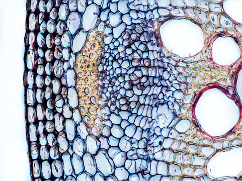
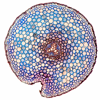
Free Images: "bestof:Tertiary Endodermis Iris florentina.png Tertiary Endodermis of Iris florentina; 1 Durchlasszelle 2 Rindenparenchym 3 Endodermis 4 Perizykel 5 Phloem 6 Xylem"
Tertiary_Endodermis_Iris_florentina.png
Root_of_Allium_cepa.png
Root_Secondary_growth.png
Racine2_Dutch_text.jpg
The_Oak_(Marshall_Ward)_Fig_5.jpg
The_Oak_(Marshall_Ward)_Fig_24.jpg
Iris_germanica_florentina_-_Köhler–s_Medizinal-Pflanzen-078.jpg
Flowers_(16264310509).jpg
The_wonderful_new_rose_hoosier_beauty_(16201164278).jpg
Autumn_bulbs_catalogue_(16451004812).jpg
Bulbs_and_plants_(16387456232).jpg
Terms of Use   Search of the Day