Click Here for More Images from iStock- 15% off with coupon 15FREEIMAGES 
The three-dimensional structure of proteins is represented in a variety of conformations. This image is based on the ribbon structure of a protein. It is suitable for biology notebooks and book covers.\nThis image has not been created by AI.
mRNA (multicoloured strand) is decoded in a ribosome to produce an aminoacid chain, that folds into a protein (red). tRNA (dark violet) molecules carry single aminoacids that are incoporated into the growing chain.
3D cartoon and Gaussian surface models, PDB 7las, chain id color scheme, white background
A model of a nucleosome. Isolated on black.
Nucleosome structure. Structure of an oligonucleosome, showing the packaging of DNA in chromosomes. 3D illustration.
Molecular Model of Human Insulin with disulfide bonds shown.
Endosomes - 3d rendered image of Endosomes are membrane-bound organelles. They are parts of endocytic membrane transport pathway.  Endomembrane system in cells. Bilayer Lipid Membrane. Abstract medical illustration. Microscopic view. Microbiologic research concept.
Rendering of cell membrane lipids
Ribosome, 3d illustration of Translation
CRISPR-Cas9 gene editing technology
The LDL-receptor (red) is a membrane protein found in almost every human cell. It binds to the apoprotein B100 from LDL-particles and mediates their internalization (endocytosis) in the cell. This occurs in almost all cells but mainly in the liver where it removes about 70 % of LDL from the circulation.
Covid-19 Coronavirus Variants Omicron Delta Gamma Beta Alpha Pandemic Virus Disease Mutation
Cyclin is a family of proteins that controls the progression of a cell through the cell cycle by activating cyclin-dependent kinase (CDK) enzymes or group of enzymes required for synthesis of cell cycle. 3D cartoon model, PDB 2i53, white background.
3d rendering of Easter eggs background
Human alcohol dehydrogenase (ADH1A) protein model
Endosomes - 3d rendered image of Endosomes are membrane-bound organelles. They are parts of endocytic membrane transport pathway.  Endomembrane system in cells. Bilayer Lipid Membrane. Abstract medical illustration. Microscopic view. Microbiologic research concept.
The crystal structure of the tumor marker protein. The 3D model of the biological macromolecule.
plant Zea Stem C.S. under light mircoscope with white background
orange: RNA primer; red: RNA template; light blue: proofreading riboexonuclease; mid blue: helicase; dark blue: RNA directed RNA polymerase; violet: non structural proteins (NSPs).
Inside protein molecule
DNA with biological concept, 3d rendering. 3D illustration.
Photomicrograph of filamentous blue-green algae, Oscillatoria species. Each band along the stand is one cell. This filament is very short probably having recently parted from a longer one. Filaments are typically many times longer. This algae is mobile, it moves and twists through the water. San Francisco Bay, California, USA. Live specimen. Wet mount, 40X objective, transmitted brightfield illumination. Note - motion blur of live specimen, very shallow depth of field, chromatic aberration and uneven focus are inherent in light microscopy.
3D cartoon model, secondary structure color scheme, PDB 1acc, white background
3d Plant Cell structure isolated on white background
The integration of fluidics and optics, antibodies molecule in flow-through nanohole arrays, has enabled increased transport of analytes to sensing surfaces 3d rendering
Bacteria pathogen - 3d rendered microbiology image. Illustrate of Lactobacillus Bulgaricus Bacteria, Microbacterium Tuberculosis Bacteria (MTB). Medical research, health-care concept. SEM (TEM) scanning view
From left to right: the NMDA and  AMPA receptors transport calcium cations into neurons after being activated by the neurotransmitter glutamate and  the GABA receptor (right) transport chloride anions after the activation by gamma aminobutyrate. PDB entries: 6wht,  3kg2, 6d6u
3D rendered part of a DNA chain on a black background with a copy space for scientific designs
Internal structure of an animal cell, 3d rendering. Section view. Computer digital drawing.
Particles, medical concept
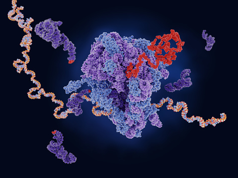
Free Images: "bestof:TRNA ribosomes diagram hy.svg en Diagram showing how proteins are produced with tRNA mRNA and ribosomes in Armenian 2016-02-03 File TRNA ribosomes diagram en"
Diagram_of_the_Federal_Government_and_American_Union_edit.jpg
Airport traffic pattern.jpg
A Lend-Lease British Valentine tank produced for the Soviet Union.jpg
Timeline energy companies NL.png
1724_De_L'Isle_Map_of_Persia_(Iran,_Iraq,_Afghanistan)_-_Geographicus_-_Persia-delisle-1724.jpg
structure-plan-coding-strategy-620304.jpg
Commodore-64-Back.jpg
Wii-Console-Back.jpg
Kotzebue, Alaska.jpg
Jerusalem panorama early twentieth century2.jpg
Kids playing and showing off newly tied knots.jpg
Tokyo hy power hl 180v 2m band rf power amplifier.jpg
Employee showing area of woodland being invaded by garlic mustard plant.jpg
Video production workers in studio studying bank of monitors showing camera views.jpg
Aerial view of tidewater glacier showing medial and lateral moraines.jpg
Fisherman and children fishing and showing huge fish catch.jpg
Son showing trout fish he caught mother stads proudly behind at lake.jpg
Side channel of the Missouri river showing erosion and downed trees.jpg
Man gives a thumbs up to visitors showing his total enjoyment with this new electric car.jpg
Male walrus out of water lifted head proudly showing their large tusks.jpg
Man proudly showing off fish that was just caught by two young boys.jpg
St Marks lighthouse stands among the trees by the blue ocean showing mariners the way.jpg
Prescribed burn of marsh grasses showing heavy plant growth with flames and smoke.jpg
Max_Brückner_-_Otto_Henning_-_Richard_Wagner_-_Final_scene_of_Götterdämmerung.jpg
Two older men enjoying fishing from boat one man is standing and raising hat while showing the fish he just caught.jpg
William_Simpson_-_George_Zobel_-_England_and_America._The_visit_of_her_majesty_Queen_Victoria_to_the_Arctic_ship_Resolute_-_December_16th,_1856.jpg
Jerusalem Oesterreichisches Hospiz.JPG
Remington M1867.jpg
World_Map_1689.JPG
Close up wall texture.jpg
Red brick detailed photo.jpg
FieldsMedalFront.jpg
Painted lady butterfly vanessa carui.jpg
White and blue stone brick wall.jpg
The_Anti-Slavery_Society_Convention,_1840_by_Benjamin_Robert_Haydon.jpg
Elizabeth_I_Steven_Van_Der_Meulen.jpg
Portrait Caliph Abdulmecid II.jpg
بيروت_القرن_١٩_أسود_أبيض.jpg
Layla_and_Majnun2.jpg
Giuseppe Verdi, Un Ballo in maschera, Vocal score frontispiece - restoration.jpg
Kamianets-Podilskyi_map_1691.jpg
Jacques-Louis David, The Coronation of Napoleon edit.jpg
Global tropical cyclone tracks-edit2.jpg
TRNA ribosomes diagram hy.svg
TRNA ribosomes diagram pl.svg
TRNA ribosomes diagram en.svg
TRNA ribosomes diagram ru.svg
Ribossomo tradução mRNA.svg
Ribosome mRNA translation zh.svg
Ribosome mRNA translation bg.svg
Ribosom mRNA translation de.svg
Ribosome mRNA translation it.svg
Ribosome mRNA translation pl.svg
Ribosome mRNA translation es.svg
Ribosome mRNA translation cs.svg
Ribosome mRNA translation tr.svg
Ribosome mRNA translation en.svg
Ribosome mRNA translation fr.svg
Zenith-Nadir-Horizon hy.svg
Average prokaryote cell- ru.svg
Average prokaryote cell- uk.svg
Prokaryote cell-ro.svg
Average prokaryote cell- nl.svg
Prokaryote cell diagram international.svg
Zelula prokarioto tipikoa.svg
Snake lemma nat2.svg
Cell membrane detailed diagram en.svg
TypesOfLenses.svg
Synergic.svg
Atmosphaerenzusammensetzung.svg
SpaceStationCycle.svg
Diagram of swine influenza symptoms EN.svg
Diagram of influenza infection symptoms EN.svg
ComputerMemoryHierarchy.svg
Machined Holes.svg
Catabolism schematic arm.svg
PD Diagram of swine flu symptoms EN.svg
Crossover genes zh.svg
Borda-repeating-circle.svg
Binary Representations.svg
Trilobite cranidium numbered.svg
Trilobite cranidium-en.svg
Gps error diagram.svg
Project-triangle.svg
Chloroplast diagram bs plain.svg
Tornado Alley Diagram fr.svg
Catalisis enzimatica1.svg
Tree secondary components diagram.svg
Mureine.svg
Acetabularia mediterranea life.svg
Blood Compatibility.svg
Trilobite cephalon areas numbered.svg
Trilobite cephalon areas-en.svg
Hardware Virtualization (copy).svg
WP Offline Proccess en.svg
Cellular tight junction-en.svg
Animal mitochondrion diagram unlabelled.svg
Fluorine shielding.svg
Cellular tight junction-it.svg
EFA to Eicosanoids ja.svg
Terms of Use   Search of the Day