Click Here for More Images from iStock- 15% off with coupon 15FREEIMAGES 
COVID-19 new mutation variant - 3d rendered image. Pandemic wave.\nViral Infection concept. Delta, Omicron (B.1.1.529), Centaurus (B.1.1.529.2.75) (BA.2.75), Aeterna (BA.4.6) variant. Sars-cov-2, 2019-nCoV, Coronavirus.\nAntibody, Antigen, stealth, Vaccine technology concept.\nB117 - COVID-19 Variant,\nSARS-CoV-2 BA.2 Variant,\nSARS-CoV-2 BA.5 Variant,\nSARS-CoV-2 Omicron Variant,\nSaRS-CoV-2 Deltacron Variant
Nodularia sp. toxic algae under microscopic view, nitrogen-fixing cyanobacteria, cyanotoxin, harmful to humans, x40
Photomicrograph of Histological analysis of Intramedullary SOL showing Astrocytoma, WHO grade-4
Macro view of isolated H1N1 swine flu virus. Clipping path included.
Alveolar soft part sarcoma, or ASPS, is a cancer that can come from different types of soft tissue, such as muscle, fat, or nerves.
Stratified flat epithelium under microscope
Epidermis. Electron microscope micrograph showing a keratinocyte of spinous layer. The epithelial cell has a polygonal shape, central nucleus with nucleolus, cytoplasm full of keratin filament bundles, and numerous dark desmosomes crossing the intercellular spaces.
corn stem micrograph with dye
The study of plant tissues under the microscope in the laboratory.
Endosomes - 3d rendered image of Endosomes are membrane-bound organelles. They are parts of endocytic membrane transport pathway.  Endomembrane system in cells. Bilayer Lipid Membrane. Abstract medical illustration. Microscopic view. Microbiologic research concept.
PaPilloma virus on a dark background. 3d illustration
plant Flower bud of Lilium brownii var.viridulum T.S.\nunder light mircoscope with white background
Scientifically accurate 3D structure of Hepatitis A Capsid based on PDB : 4QPI.
Burkitt lymphoma is a form of non-Hodgkin's lymphoma in which cancer starts in immune cells called B-cells. Recognized as a fast growing human tumor, Burkitt lymphoma is associated with impaired immunity and is rapidly fatal if left untreated.
COVID-19 Corona virus with spike glycoprotein are floating on the air . Blue color background . 3D rendering .
Looking through a window underwater where limpets and saline have grown over window.
3d rendering of a virus
Quantum Gravity 3d rendered image of a comprehensive exploration of Quantum Gravity theories. String theory, loop quantum gravity (LOG), causal dynamical triangulation, twistor theory, gravitational waves, spacetime, gravity waves, quantum mechanics, quantum entanglement concepts.
Background of mold in the wood of the house.
Liver Fluke Section under light microscope with white background
Toxoplasma gondii oocyst under the microscope, isolated
Blastomycosis is a fungal infection caused by inhaling Blastomyces dermatitidis spores. If it involves only the lungs, it is called pulmonary blastomycosis. Only about half of people with the disease have symptoms, which can include fever, cough, night sweats, muscle pains, weight loss, chest pain, and feeling tired.
Thyroid cancer: Microscopic image of Metastatic papillary carcinoma of thyroid. Reactive change. Lymph node carcinoma.
Conceptual visualization of the swine influenza virus (h1n1)
Nucleosome, molecular model. 3D illustration. The nucleosome consists of a DNA double helix wrapped around a core of histone proteins.
COVID-19 new Omicron sub-variant BQ.1.1 - 3d rendered image. Pandemic wave.\nViral Infection concept. Delta, Omicron (B.1.1.529), Centaurus (B.1.1.529.2.75) (BA.2.75), Aeterna (BA.4.6) variant. Sars-cov-2, 2019-nCoV, Coronavirus.\nAntibody, Antigen, stealth, Vaccine technology concept.\nB117 - COVID-19 Variant,\nSARS-CoV-2 BA.2 Variant,\nSARS-CoV-2 BA.5 Variant,\nSARS-CoV-2 Omicron Variant,\nSaRS-CoV-2 Deltacron Variant
False colour transmission electron microscope (TEM). Neuron cell body: nucleus (magenta), mitochondria (green), lysosomes (dark blue), multivesicular bodies (red), RER (brown) and Golgi (yellow).
Abstract marble texture background stock photo, suitable for design.
microscopic view of plant cells for botanic education and analysis
Mesenchymal dysplasia. Site: Placenta
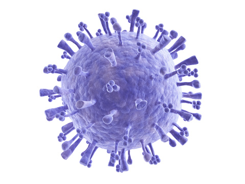
Free Images: "bestof:TEM phil CDC 11635 lores.jpg en This colorized transmission electron micrograph TEM revealed the presence of a number of Novel H1N1 virus virions in this tissue"
Ebola_Virus_Particles.jpg
Religión en Isla Margarita, Valle del Espíritu Santo.jpg
Pieter_Brueghel_the_Elder_-_The_Dutch_Proverbs_-_Google_Art_Project.jpg
Sarcomere schema et photo-FR.svg
"Carretelas_en_la_Vega".jpg
El_sueño_de_San_José_(Museo_del_Prado).jpg
Diagrama enclave exclave.svg
Swine flu infection exponent by county June 2009.svg
Enterobius_vermicularis_LifeCycle_B.svg
Sebastião_de_Portugal,_c._1571-1574_-_Cristóvão_de_Morais.png
TEM_phil_CDC_11635_lores.jpg
Nipah.jpg
H5N1_virions_108,000x.jpg
B00528-Swine-flu.png
A_(H5N1)_virion,_a_type_of_bird_flu_virus_which_is_a_subtype_of_avian_influenza_A.jpg
Chikungunya_virus_particles-PHIL-17369.jpg
Poxvirus_of_molluscum_contagiosum_TEM_B81-166_lores.jpg
Rotavirus_TEM_B82-0337_lores.jpg
Scanning_Electron_Micrograph_of_a_Flea.jpg
Smallpox virus virions TEM PHIL 1849 (crop).jpg
H1N1_virus_particles.jpg
H1N1_virus_particles_II.jpg
Dengue.jpg
Electron_Micrograph_of_Flea_derivate.jpg
Coronavirus.jpg
LegionellaPneumophila.jpg
Giardia lamblia.bmp.jpg
Campylobacter_jejuni_5778_lores.jpg
Streptococcus_pneumoniae-263.jpg
TEM of L-form bacteria-Mark Leaver Newcastle University.tif
TEM of L-form bacteria-Mark Leaver Newcastle University.jpeg
Plasmodium_falciparum_rings_form_parasites4885_lores.jpg
Bourbon_virus_endocytosis_(EID_2015_Fig_2d).jpg
Multidrug-resistant Klebsiella pneumoniaeand neutrophil.bmp.jpg
Cygnet_River_square_(EID_2012_Fig1a).jpg
Blood clot in scanning electron microscopy.jpg
Giardia_1.jpg
MRSA_SEM_7821_lores.jpg
Human skeletal muscle tissue 1 - TEM.jpg
Human skeletal muscle tissue 2 - TEM.jpg
Human skeletal muscle tissue 3 - TEM.jpg
Eosinophil TEM.jpg
Mitochondria - TEM.jpg
Norovirus_EM_PHIL_2172_lores.jpg
H1N1_influenza_virus.jpg
Pancreatic cells - TEM.jpg
SARS_CoV.jpg
Malassezia_furfur_SEM_lores.jpg
Banna_virus_EM.jpg
Mitochondria, mammalian lung - TEM (2).jpg
Mitochondria, mammalian lung - TEM.jpg
Clara cell lung - TEM.jpg
Pancreatic acinar cells - TEM.jpg
A red blood cell in a capillary, pancreatic tissue - TEM.jpg
Desmosome - epithelial cell from mammalian lung tissue - TEM.jpg
Histopathologic_features_of_aspergillosis_including_the_presence_of_conidial_heads_PHIL_4335_lores.jpg
TEM of silicon nanowire (5887830328).jpg
Fibers of Collagen Type I - TEM .jpg
Macrophage in the alveolus Lung - TEM.jpg
Fibers of Collagen Type I - TEM.jpg
Spindle centriole - embryonic brain mouse - TEM.jpg
Deinococcus_radiodurans.jpg
Staph_aureus_outside_a_white_blood_cell.jpg
Transmission Electron Microscope Kemira.jpg
Wangiella_dermatitidis_PAS_stain_PHIL_3781_lores.jpg
Neodymium Crystal Structure Nd2Fe14B.jpg
Retino ME con sezioni.jpg
25K15pA9Def4sec Arman 4 Box1.png
Terminal_chlamydospores_of_the_fungus_Microsporum_audouinii_PHIL_3054_lores.jpg
CDC_MMWR_M8d0430a2f.png
Golgi in the cytoplasm of a macrophage in the alveolus (lung) - TEM.jpg
Junctional complex and pinocytotic vesicles - embryonic brain-TEM.jpg
Unctional complex and pinocytotic vesicles - embryonic brain - TEM.jpg
Cilia.jpg
Embryonated_chicken_egg-10759_lores.jpg
Bourbon_virus_sphere_(EID_2015_Fig_2b).jpg
Bourbon_virus_filament_(EID_2015_Fig_2a).jpg
Countries with confirm double cases of swine flu.png
Mammalian pancreatic tissue, nuclear pores and rough endoplasmic reticulum.jpg
Velvet_ant_head_anterior_--_SEM_photo.jpg
Taenia_solium_tapeworm_proglottids_5261_lores.jpg
Velvet_ant_stinger_(tip)--scanning_electron_microscope_image.jpg
Bourbon_virus_filament_crop_(EID_2015_Fig_2a).jpg
Bourbon_virus_sphere_crop_(EID_2015_Fig_2b).jpg
Schistosoma_haematobium_egg_4842_lores.jpg
Gravid_proglottid_from_the_cestode_Taenia_saginata_5259_lores.jpg
7175_lores.jpg
7176_lores.jpg
Bacillus subtilis.jpg
Cygnet_River_phylogeny_(EID_2012_Fig2).jpg
Quatrain_for_a_King_WDL7028.png
A_New_Compilation_of_Events_from_the_Xuanhe_Period_WDL7101.jpg
Idylls_WDL10691.jpg
Two desmosomes - TEM.jpg
Chlamydomonas TEM 01.jpg
Chlamydomonas TEM 02.jpg
Chlamydomonas TEM 03.jpg
Chlamydomonas TEM 04.jpg
Chlamydomonas TEM 05.jpg
Terms of Use   Search of the Day