Click Here for More Images from iStock- 15% off with coupon 15FREEIMAGES 
The study of plant tissues under the microscope in the laboratory.
Microscopic image of the cross section of the root of a buttercup (crowfoot) plant - Ranunculus repens. The propeller shaped pattern in the center is the vascular tissue for transporting water and nutrients up and down the plant. The circles are the individual cells.
The xylem and phloem that make up the vascular tissue of the stem are arranged in distinct strands called vascular bundles, which run up and down the length of the stem
plant Stem-Collenchyma C.S. under light mircoscope with white background
Blue colored stem cells of a lentil plant under the microscope.
Blastomycosis is a fungal infection caused by inhaling Blastomyces dermatitidis spores. If it involves only the lungs, it is called pulmonary blastomycosis. Only about half of people with the disease have symptoms, which can include fever, cough, night sweats, muscle pains, weight loss, chest pain, and feeling tired.
Cotton plant root, under the light microscope. Longitudinal section through the root of Gossypium hirsutum, upland or also Mexican cotton. Light micrograph at 20X magnification. Isolated, over white.
corn stem micrograph with dye
Epidermis. Electron microscope micrograph showing a keratinocyte of spinous layer. The epithelial cell has a polygonal shape, central nucleus with nucleolus, cytoplasm full of keratin filament bundles, and numerous dark desmosomes crossing the intercellular spaces.
Particles, medical concept
Cross-section Plant Stem under the microscope for classroom education.
plant rape flower cross section under light mircoscope with white background
Cross section leaf of plant under the light microscope view for botany education.
Diffuse large B-cell lymphoma, or DLBCL, is a cancer that starts in white blood cells called lymphocytes.
Lime trunk
Macro photography of Crystal texture background, nature pattern.
Leiden leaf c.s. under microscope
Photomicrograph of flatworm,
Plant tissue Structure, section (tissue) of stem plant tissue under a light microscope.
Nymph T.S. under light microscope with white background
Microscopic fungi Malassezia furfur, showing yeast cells and hyphae. dermatophytes, Nail scraping or skin scraping for fungus test in microbiology laboratory.
Microscopic image of dog stomach cardiac region
linden stem (Tilia platyphyllos) cross section under the microscope showing phloem, vascular cambium, medullary ray and pith - optical microscope x32 magnification
Paraganglioma is a type of neuroendocrine tumor that forms near certain blood vessels and nerves outside of the adrenal glands. The adrenal glands are important for making hormones that control many functions in the body and are located on top of the kidneys.
Microscope photo of a sambucus stem with parenchyma cells.
Micro world
Host cells with spores (mold) are inside wood under the microscope for education.
Photomicrograph of
Aerial view of loyster farm by Ardara, County Donegal - Ireland.
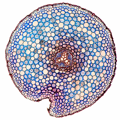
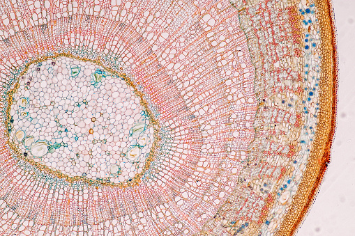
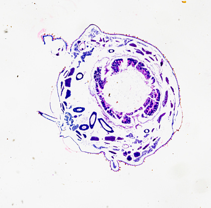
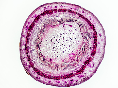
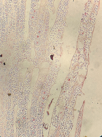
Free Images: "bestof:Stem-histology-cross-section-tag.svg Flax stem cross-section Legend pith protoxylem xylem II phloem I Sclerenchyma bast fibre cortex epidermis Coupe"
Stem-histology-cross-section-tag.svg
Labeledstemforposter copy new.jpg
The_Oak_(Marshall_Ward)_Fig_12.jpg
Root_of_Allium_cepa.png
The_Oak_(Marshall_Ward)_Fig_5.jpg
Leaf anatomy universal.png
The_Oak_(Marshall_Ward)_Fig_17.jpg
The_Oak_(Marshall_Ward)_Fig_24.jpg
The_Oak_(Marshall_Ward)_Fig_9.jpg
Image_from_page_236_of_"The_structure_and_development_of_mosses_and_ferns_(Archegoniatae)"_(1918).jpg
Terms of Use   Search of the Day