Click Here for More Images from iStock- 15% off with coupon 15FREEIMAGES 
Backgrounds of Bacteria and Mold under the microscope in Lab.
Hospital
Histopathology of diffuse sclerosing glomerulonephritis, light micrograph, photo under microscope
Serous carcinoma is the most common type of ovarian cancer, accounting for approximately 75% of epithelial ovarian cancers.
Colored plant cells with damages caused by a parasitic animal under the microscope.
Male Caucasian dentist, tutoring the female student in his private ordination, explaining her the x-ray image
PAPS Smear study of a young women under microscopy showing Atrophic changes in uterus which is unusual at young age.
Thyroid cancer: Microscopic image of Metastatic papillary carcinoma of thyroid. Reactive change. Lymph node carcinoma.
Acute ordinary hepatitis liver biopsy under light microscopy
Showing Light micrograph Type of Tissue Human under the microscope in Lab.
Mesenchymal dysplasia. Site: Placenta
Breast tissue adenocarcinoma immunohistochemistry. Estrogen receptors.
Doctor showing X rays to his patient
Gynecologist doctor discussing baby sonography image to young pregnant lady during meeting in clinic, doc consulting expectant woman in hospital
Spleen Tissue From Dog seen on microscope at 200x Magnification. Phase Contrast Optical Microscope
Photomicrograph of Stomach Adenocarcinoma. Gastric Adenocarcinoma.
Histological Cerebellum human, Sympathetic ganglion human And Spinal cord human under the microscope for education.
Carcinoid tumors are a type of slow-growing cancer that can arise in several places throughout your body. Carcinoid tumors, which are one subset of tumors called neuroendocrine tumors, usually begin in the digestive tract (stomach, appendix, small intestine, colon, rectum) or in the lungs. Site: Ovary
Kidney cancer: Microscopic image of metastatic clear cell carcinoma of kidney, the most common type of renal cell carcinoma. Show brain tissue of malignant neoplasm, large atypical bizarre cells.
human Miliary tuberculosis biopsy slide under microscope zoom in different areas
Duplex sonography of human carotid artery an thyroid gland
\
Photomicrograph of Histological analysis of Intramedullary SOL showing Astrocytoma, WHO grade-4
Microscopic image of Microscopic image of nymphaea of aqustio stem
Hyaline cartilage, Elastic cartilage and Bone Human under the microscope in Lab.
Squamous cell carcinoma of the lung is a type of lung cancer. It occurs when abnormal lung cells multiply out of control and form a tumor.
Light microscope micrograph showing mitosis in onion cells of the root meristem. At bottom, is a cell in anaphase. On the top right corner, a prophase.
smoke lung with black deposit in microscopy
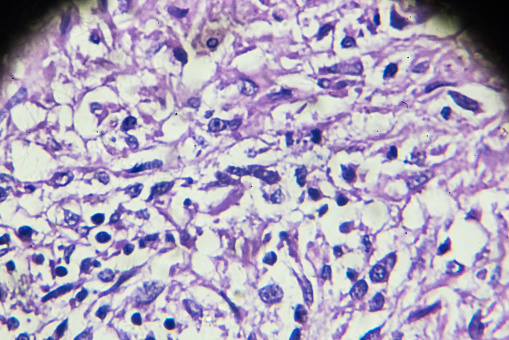
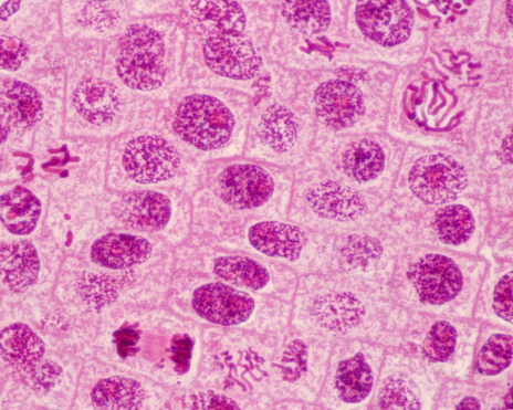
Free Images: "bestof:Squamous cell carcinoma.jpg I am a doctor and I took this picture a couples of month back in the clinic I have blocked out the patient's identity for privacy"
x-ray-medical-broken-arm-doctor-1273382.jpg
Manzanar_Relocation_Center,_Manzanar,_California._Doctor_Takahashi,_Eye,_Ear,_Nose_and_Throat_Speci_._._._-_NARA_-_538147.jpg
Squamous cell carcinoma.jpg
Squamous_Cell_Carcinoma.jpg
Squamous_Cell_Carcinoma1.jpg
Flickr_-_Official_U.S._Navy_Imagery_-_Navy_eye_doctor_examines_patient_in_Peru.jpg
Jerome_Relocation_Center,_Denson,_Arkansas._A_patient,_teacher_of_high_school_science,_being_examin_._._._-_NARA_-_538864.tif
Jerome_Relocation_Center,_Denson,_Arkansas._A_patient,_teacher_of_high_school_science,_being_examin_._._._-_NARA_-_538864.jpg
Minidoka_Relocation_Center._Doctor_Robert_H._Shiomi,_Miyoko_Shitamae,_nurse,_Jimmie_Shimizu,_patien_._._._-_NARA_-_536596.tif
Minidoka_Relocation_Center._Doctor_Robert_H._Shiomi,_Miyoko_Shitamae,_nurse,_Jimmie_Shimizu,_patien_._._._-_NARA_-_536596.jpg
Manzanar_Relocation_Center,_Manzanar,_California._Doctor_Takahashi,_Eye,_Ear,_Nose_and_Throat_Speci_._._._-_NARA_-_538147.tif
Possibly_Margaret_More,_daughter_of_Sir_Thomas_More.jpg
Terms of Use   Search of the Day