Click Here for More Images from iStock- 15% off with coupon 15FREEIMAGES 
the head of Aelurostrongylus abstrusus, cat lungworm under the microscope
Liver Fluke Section under light microscope with white background
Demodex folliculorum - parasitic mite on the eyelashes of a human eye, microscope
Blastomycosis is a fungal infection caused by inhaling Blastomyces dermatitidis spores. If it involves only the lungs, it is called pulmonary blastomycosis. Only about half of people with the disease have symptoms, which can include fever, cough, night sweats, muscle pains, weight loss, chest pain, and feeling tired.
amazing inhabitants of the microworld under a microscope
Photomicrograph of two rotifers. Rapidly rotating cilia at top of heads, internal organs visible. Live specimen. Wet mount, 10X objective, transmitted brightfield illumination.
Microscopic photo of a thin section of calcareous tufa of Holocene age.
Natural agate surfaces, suitable for backgrounds and wallpapers.
Planarian parasite (flatworm) under microscope view.
plant Zea Stem C.S. under light mircoscope with white background
Cross section of Escherichia coli bacteria
Trematode parasite under a microscope
Daphnia - micro organism
Urothelial carcinoma, high grade.  High-grade tumors have an aggressive appearance under a microscope and are assumed invasive in the kidney or ureter.
Photomicrograph of filamentous blue-green algae, Oscillatoria species. Each band along the stand is one cell. This filament is very short probably having recently parted from a longer one. Filaments are typically many times longer. This algae is mobile, it moves and twists through the water. San Francisco Bay, California, USA. Live specimen. Wet mount, 40X objective, transmitted brightfield illumination. Note - motion blur of live specimen, very shallow depth of field, chromatic aberration and uneven focus are inherent in light microscopy.
centropyxis aculeata (amoeboid organism) under the microscope - optical microscope x400 magnification
Planarian parasite (flatworm) under microscope view.
European seabass raw fish fillet for advertisement
newly hatched freshwater fish under a light microscope, isolated on white
schistosoma japonicum cercariae under light microscopy
Freshwater aquatic zooplankton and algae under microscope view
Bone marrow plasma cells .Plasma cell neoplasms are diseases in which the body makes too many plasma cells. Plasma cell neoplasms can be benign or malignant. Diagnosed with Immunostain CD138 positive.
Marine flatworm - Planaria, crawling on the glass, Black Sea
Haliotis kamtschatkana, northern abalone or pinto abalone, is a species of large sea snail, a marine gastropod mollusc in the family Haliotidae, the abalones. It has been listed as \
Eggs of helminth and Finding parasites in feces, analyze by microscope in laboratory.
Amazing Colorful Agate Crystal stone cross texture background.
Body of cabbage larvae filled with parasitic entomopathogenic nematodes -Steinernema sp.
Fresh fish  croaker on white background
Microscopic view of a cat flea eggs
Plasmacytoid urothelial carcinoma (PUC) is a unique variant of invasive urothelial carcinoma characterized by tumor cells that exhibit a striking resemblance to plasma cells, and it is known to have an aggressive behavior.
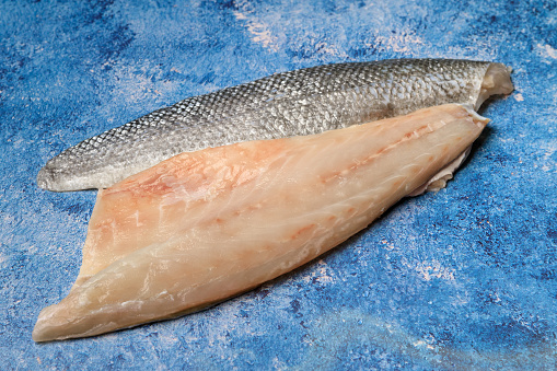
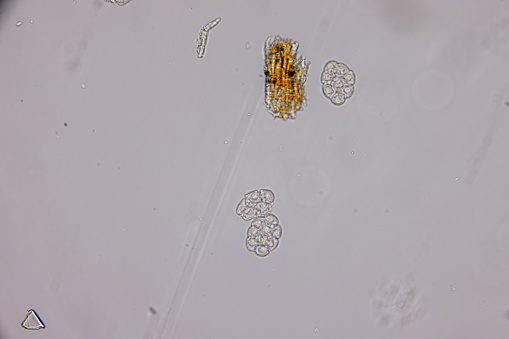
Free Images: "bestof:Solea vulgaris - Lower (left) side of the Common Sole Two specimens of the parasite Plyllonella soleae are shown on the skin.jpeg check categories 22 August"
Terms of Use   Search of the Day