Click Here for More Images from iStock- 15% off with coupon 15FREEIMAGES 
DNA sequencing blueprint RNA sequencing DNA computational models, genome helix background, gene CRISPR helix
DNA Molecule: a 3D illustration of the double helix structure reflecting the genetic code. This 3D illustration is a visualization of the DNA molecule showing its characteristic double helix structure. The model reflects the major components of DNA such as the nitrogenous bases (adenine, thymine, guanine, and cytosine), sugars, and phosphate groups that together form the genetic code. This illustration can be used for educational purposes, scientific publications or medical advertising to demonstrate the complexity and beauty of the molecule responsible for heredity and the development of life on Earth.
A plasmid is a small circular DNA molecule found in bacteria and some other microscopic organisms 3d rendering
Digital screen with DNA strands and data background. Double helix structure. Nucleic acid sequence. Genetic research. 3d illustration.
3D cartoon model, PDB 5wn4, white background
Photomicrograph of filamentous blue-green algae, Oscillatoria species. Each band along the stand is one cell. This filament is very short probably having recently parted from a longer one. Filaments are typically many times longer. This algae is mobile, it moves and twists through the water. San Francisco Bay, California, USA. Live specimen. Wet mount, 40X objective, transmitted brightfield illumination. Note - motion blur of live specimen, very shallow depth of field, chromatic aberration and uneven focus are inherent in light microscopy.
RNA And DNA concept as a deoxyribonucleic acid or a ribonucleic acid as biological molecules as a symbol of the evolution of life and genetic sequencing.
DNA and RNA Editing Concept. 3D Illustration
The Raf/MEK/ERK (MAPK) propagates growth signals in the cell. Misregulation can lead to cancer.\nThe red dots on the active EGFR are phosphorylated tyrosines. PDB entries: 1egf, 1nql, 1m17, 2jwa, 3njp, 2gs6, 1gri, 1xd2, 3ksy, 5p21, 6xi7, 6q0j, 2y4i,
plant Zea Stem C.S. under light mircoscope with white background
Diatoms, algae under microscopic view, phytoplankton, fossils, silica, golden yellow algae
3D rendered part of a DNA chain on a black background with a copy space for scientific designs
DNA structure - Deoxyribonucleic acid. 3D for medical scientific RNA research and biology genetic molecular laboratory study.
3D Render of right-handed DNA molecular structures against a white background
Genetic analysis concept. DNA molecule and genome sequence in background. 3D rendered illustration.
3d Render DNA Molecule Structure & Red, Science and Medicine Concept, Depth Of Field
Abstract DNA spiral model (done in 3d)
Myxoid liposarcoma.  Myxoid/round cell liposarcoma, or MRCLS, is one of several types of liposarcoma. Liposarcoma is a rare cancer that grows in the cells that store fat in the body. MRCLS usually grows in the arms and legs. These tumors grow slowly, and they can spread to other parts of the body
Backgrounds of Bacteria and Mold under the microscope in Lab.
Computational tomography image of the abdominal and pelvic regions.
Molecule with water background, 3d rendering. Digital drawing.
Photomicrograph of two rotifers. Rapidly rotating cilia at top of heads, internal organs visible. Live specimen. Wet mount, 10X objective, transmitted brightfield illumination.
DNA digital data storage is the process of encoding and decoding binary data to and from synthesized strands of DNA 3d rendered
DNA. Abstract 3d polygonal wireframe DNA molecule. Medical science, genetic biotechnology, chemistry biology, gene cell concept
Nodularia sp. toxic algae under microscopic view, nitrogen-fixing cyanobacteria, cyanotoxin, harmful to humans, x40
DNA and biotechnology concept
Computer generated 3D model camera positioned from all 6 directions white background with alpha channel for transparency
Fatty liver disease (steatosis) is a common condition caused by having too much fat build up in your liver. A healthy liver contains a small amount of fat. It becomes a problem when fat reaches 5% to 10% of your liver's weight.
Secreted by Staphylococcus aureus, this toxin binds with the outer membrane, with subsequent oligomerization of the toxin monomers to form water-filled channels. These are responsible for osmotic phenomena, cell depolarization and loss of vital molecules (v.gr. ATP), leading to cell death. 3D cartoon model in two purpendicular projections, chain id color scheme, based on PDB 3anz, white background.
DNA strands are arranged next to each other with decreasing size on a dark background - 3d images of dna molecules on black background, science nanotechnology, medical concept, on dark bg, hologram view.
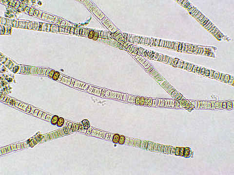
Free Images: "bestof:Single Cell Genome Sequencing (MDA).JPG en MDA Own Qianlim Unidentified biology diagrams Genome sequencing Diagrams in English"
8-cell-simple.gif
Naked human male body front anterior.png
CMB Timeline75 (Arabic Vision).jpg
Metamorphosis frog Meyers.png
Pluto System (french).jpg
Snells law wavefronts.gif
A_Specimen_by_William_Caslon.jpg
Plant cell wall diagram-en.svg
Study-flow (zh-cn).svg
Study-flow.svg
Scheme secundary active transport-en.svg
Scheme facilitated diffusion in cell membrane-en.svg
Scheme simple diffusion in cell membrane-en.svg
Scheme sodium-potassium pump-en.svg
Average prokaryote cell- en.svg
B_cell_activation_-_ru.svg
Supported bilayer.svg
Lipid vesicles.svg
Average prokaryote cell numbered.svg
Average prokaryote cell- da.svg
Beta oksydacja.svg
Black lipid membrane.svg
TRNA ribosomes diagram en.svg
Lipid bilayer fusion.svg
Endomembrane system diagram en (edit).svg
Animal cell structure en.svg
Organization levels mouse.svg
Animal cell structure fr.svg
Exocytosis types.svg
Avocado seed diagram-en.svg
Chromosome fr.svg
Semnalizare engl.svg
Single side platform diagram.svg
Pore schematic.svg
Scheme simple diffusion in cell membrane-es.svg
Average prokaryote cell- fr.svg
Scheme facilitated diffusion in cell membrane-eo.svg
Desmosome cell junction en.svg
Mature flower diagram bg.svg
Animal cell cycle-en.svg
Scheme sodium-potassium pump-en-2.svg
Plastids types en.svg
Pinozytose.svg
Innate_Immune_cells_-_ru.svg
Cell membrane detailed diagram en.svg
Tipica cellula procariote.svg
Gap cell junction-en.svg
Scheme simple diffusion in cell membrane-de.svg
Complete neuron cell diagram ca.svg
Complete neuron cell diagram en.svg
Plant cell structure-en.svg
Animal mitochondrion diagram en (edit).svg
Ribosome mRNA translation en.svg
Cellular tight junction-en.svg
Scheme secundary active transport-pl.svg
ShockleyQueisserVOC.svg
ShockleyQueisserZoomedIn.svg
Biological classification L Pengo.svg
ShockleyQueisserFullCurve.svg
Animal mitochondrion diagram en.svg
Animal Cell.svg
Main protein structure levels en.svg
Eukaryotic cilium diagram en.svg
Complement pathway.svg
Structure varna.svg
Cyanogene Glukoside.svg
Hdl2.svg
Launch Wave3.svg
Fuel cell EN.svg
Launch Wave2.svg
Statics.svg
Zellatmung-uebersicht.svg
LePUS3 Vocabulary.svg
Serpent.svg
Prim sprossachse.svg
Funcmap ajl.svg
Intermediate filament-HE.svg
Tympanic membrane.svg
Scheme pgp.svg
Road-sign-0036.svg
Esitumallinen solu.svg
Water lock door principle.svg
Cancer stem cells text resized.svg
Hdl1.svg
Furaptra.svg
Konische Trommelförderung.svg
MinCDE System.svg
Gap cell junction zh-cn.svg
Prokaryote cell diagram de.svg
RFLP genotyping gl.svg
Rank product.svg
Isogamy.svg
Rotation Nav.svg
NACA0015 a.svg
Diagrama d'una cèl·lula procariota.svg
Average prokaryote cell-pl.svg
Endosymbiosis.svg
CD8 receptor.svg
Cyanogene Glukoside-fr.svg
Achromat pl.svg
Terms of Use   Search of the Day