Click Here for More Images from iStock- 15% off with coupon 15FREEIMAGES 
Epidermis. Electron microscope micrograph showing a keratinocyte of spinous layer. The epithelial cell has a polygonal shape, central nucleus with nucleolus, cytoplasm full of keratin filament bundles, and numerous dark desmosomes crossing the intercellular spaces.
plant Zea Stem C.S. under light mircoscope with white background
Toxocara canis second stage larvae hatch from eggs in microscope. Toxocariasis, also known as Roundworm Infection, causes disease in humans
Blastomycosis is a fungal infection caused by inhaling Blastomyces dermatitidis spores. If it involves only the lungs, it is called pulmonary blastomycosis. Only about half of people with the disease have symptoms, which can include fever, cough, night sweats, muscle pains, weight loss, chest pain, and feeling tired.
Microscopic photo of a thin section of calcareous tufa of Holocene age.
Photomicrograph of filamentous blue-green algae, Oscillatoria species. Each band along the stand is one cell. This filament is very short probably having recently parted from a longer one. Filaments are typically many times longer. This algae is mobile, it moves and twists through the water. San Francisco Bay, California, USA. Live specimen. Wet mount, 40X objective, transmitted brightfield illumination. Note - motion blur of live specimen, very shallow depth of field, chromatic aberration and uneven focus are inherent in light microscopy.
Freshwater Closterium algae (unicellular charophyte green algae) - optical microscope x200 magnification
Close up of small feeding tick with shiny pinhead. Nikon D7200 with Nikon 105mm macro lens & extension tube, SB 21 flash. Photo taken in Gainesville, Florida
the tail of Aelurostrongylus abstrusus, cat lungworm under the microscope
Particles, medical concept
Scalp and hair follicles of human under the microscope in Lab.
Diffuse large B-cell lymphoma, or DLBCL, is a cancer that starts in white blood cells called lymphocytes.
Green algae cells background
Liver Fluke Section under light microscope with white background
Microscope photo of a sambucus stem with parenchyma cells.
Trematode parasite under a microscope
Leiden leaf c.s. under microscope
Natural agate surfaces, suitable for backgrounds and wallpapers.
Enlarge microscopic view of larvae stage of crustacean
Structure of Escherichia Coli
Microscopic view of Salmonella enterica serotype Typhi that causes typhoid fever. 3D illustration
Photomicrograph of two rotifers. Rapidly rotating cilia at top of heads, internal organs visible. Live specimen. Wet mount, 10X objective, transmitted brightfield illumination.
Epidermis. Electron microscope micrograph of an epidermis spinous layer. The keratinocytes show polygonal shapes, central nucleus with nucleolus, cytoplasm full of keratin filament bundles, and numerous desmosomes crossing the intercellular spaces.
Lymphocytic myocarditis. Myocarditis is an inflammation of the heart muscle. Lymphocytic refers to the type of cell seen in the affected tissue. A lymphocyte is a type of immune function cell.
Human hair with dandruff and close-up view of microscopic fungi Malassezia furfur that cause dandruff and seborrhoeic dermatitis, 3D illustration
plant rape flower cross section under light mircoscope with white background
Lily anther c.s. under microscope
stx2a is a subtype of Shiga toxin-producing E. coli (STEC) linked to Hemolytic Uremic Syndrome (HUS), a severe condition impacting kidney function and blood clotting. STEC poses a significant risk of foodborne illness and can spread within communities including family members.
microscopy micrograph animal, conjugation of Paramecium caudatum, magnification 200X
Monkeypox Virus. 3D Render
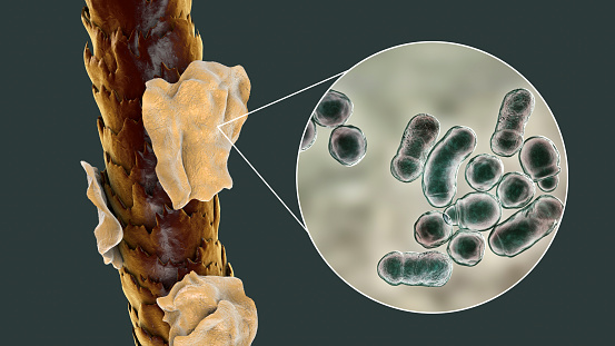
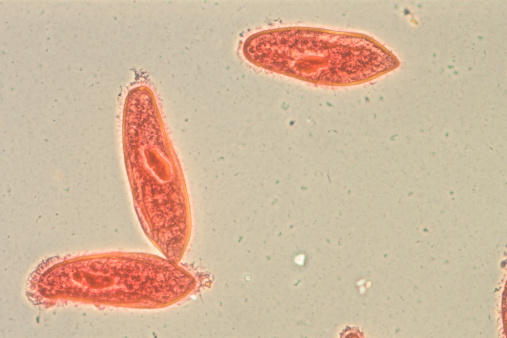
Free Images: "bestof:Schistosome Parasite SEM.jpg This schistosome parasite enters the body through the skin of persons coming in contact with infested waters The adult worm lives"
Triepeolus_lunatus,_f,_back,_Charleston_Co.,_SC_2016-09-08-18.14.jpg
Triepeolus_lunatus,_f,_right_side,_Charleston_Co.,_SC_2016-09-08-18.37.jpg
Triepeolus_lunatus,_f,_face,_Charleston_Co.,_SC_2016-09-08-18.23.jpg
Haeckel_Batrachia.jpg
Design_Analysis_of_the_P-38_Lightning.png
Symptoms of Raw fish infection.svg
Side effects of alprazolam.svg
Design_Analysis_of_the_Boeing_B-17G_Flying_Fortress.png
Schistosome_Parasite_SEM.jpg
Cercarial_LifeCycle.png
schistosome_parasite.png
FMIB_51770_Head_of_adult_Trout-worm,_Dib_thrium_e_rdiceps,_a_parasite_of_Salmo_clarkii_From_intestine_of_white_pelican,_Yellowstone_Lake.jpeg
Schistosoma 20041-300.jpg
Ringworm_due_to_Trichophyton_rubrum_PHIL_2909_lores.jpg
Salmon.jpg
Symptoms of Raw fish infection.png
Haeckel_frogs_with_labels.jpg
Scabies_LifeCycle(French_version).GIF
Ganges_River_at_Haridwar_-_1858.jpg
Haridwar,_Gateway_to_Hari,_19th_century.jpg
Terms of Use   Search of the Day