Click Here for More Images from iStock- 15% off with coupon 15FREEIMAGES 
Blue colored stem cells of a lentil plant under the microscope.
Colorful seamless pattern with Crystal stones.
abstract binary random white background 3D renderingabstract background 3D rendering - letters conceptabstract background 3D rendering -  multi ethnic conceptabstract background 3D rendering - letters concept
Particles, medical concept
Histological Pancreas human, Liver human, Vermiform appendix human and Kidney Human under the microscope for education.
corn stem micrograph with dye
Epidermis. Electron microscope micrograph showing a keratinocyte of spinous layer. The epithelial cell has a polygonal shape, central nucleus with nucleolus, cytoplasm full of keratin filament bundles, and numerous dark desmosomes crossing the intercellular spaces.
Blastomycosis is a fungal infection caused by inhaling Blastomyces dermatitidis spores. If it involves only the lungs, it is called pulmonary blastomycosis. Only about half of people with the disease have symptoms, which can include fever, cough, night sweats, muscle pains, weight loss, chest pain, and feeling tired.
Kidney cancer: Clear cell carcinoma. fuhrman nuclear grade-II, Renal growth shows a malignant tumor, renal cell carcinoma in between renal tubules.
abstract alloy texture design background.
Cross sections of the plant root under the microscope view for education
Pine Root C.S. under light mircoscope with white background
Microscope photo of a sambucus stem with parenchyma cells.
Close-up shooting of a green agate stone mineral surface isolated on white background. Its a macro shot shows nature beautiful details with back lit. Agate is a kind of natural gemstone. Many vibrant colors turquoise, orange, blue, yellow, green. Abstract pattern. Good for Wallpaper texture background. Orange.
Colorful, abstract micrograph of a fish scale, calico bass,Paralabrax clathratus, with polarization at 40x.
Diffuse large B-cell lymphoma, or DLBCL, is a cancer that starts in white blood cells called lymphocytes.
microscopy micrograph plant tissue, stem of pumpkin, magnification 200X
A model of the phospholipid bilayer structure that makes up living cells and their contents. Its raw, beautiful appearance evokes images of biology and biochemistry. It can be used in magazines and textbooks and on the covers of presentations.\nThis image was not created by AI.
Light micrograph of autonomic sympathetic ganglion stained with Cajals silver nitrate showing a mixture of multipolar neuronal cell bodies and nerve fibres.
Micro world
The study of plant tissues under the microscope in the laboratory.
Microscopic photo of a thin section of calcareous tufa of Holocene age.
Neuroendocrine carcinoma well differentiated (carcinoid) rectum with immunostain synaptophysin
Kidney cancer: Microscopic image of metastatic clear cell carcinoma of kidney, the most common type of renal cell carcinoma. Show brain tissue of malignant neoplasm, large atypical bizarre cells.
Close-up shooting of a green agate stone mineral surface isolated on white background. Its a macro shot shows nature beautiful details with back lit. Agate is a kind of natural gemstone. Abstract pattern. Good for Wallpaper texture background. Turquoise, blue
Reticular cell is a type of fibroblast. They are found in many tissues including the spleen, lymph nodes and lymph nodules. Pathology of human.
Close up of texture and colors of the bottom of a burnt pot.
Stratified flat epithelium under microscope
Yellow abstract background
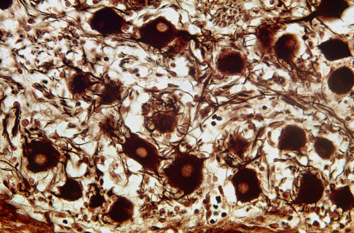
Free Images: "bestof:Schistosoma haematobium egg 4842 lores.jpg This micrograph reveals an egg from the trematode Schistosoma haematobium Schistosoma haematobium is one of the"
Schistosoma life cycle.svg
Schistosoma_haematobium_egg_4842_lores.jpg
Schistosoma_mansoni_egg_4841_lores.jpg
S_interculatum_eggS.jpg
S_mekongi_eggR.jpg
Schistosoma mansoni2.jpg
Schistosoma 20041-300.jpg
Trematode lifecycle stages.png
Giardia_1.jpg
Terms of Use   Search of the Day