Click Here for More Images from iStock- 15% off with coupon 15FREEIMAGES 
Toxocara canis second stage larvae hatch from eggs in microscope. Toxocariasis, also known as Roundworm Infection, causes disease in humans
plant Zea Stem C.S. under light mircoscope with white background
Microscopic photo of a thin section of calcareous tufa of Holocene age.
Blastomycosis is a fungal infection caused by inhaling Blastomyces dermatitidis spores. If it involves only the lungs, it is called pulmonary blastomycosis. Only about half of people with the disease have symptoms, which can include fever, cough, night sweats, muscle pains, weight loss, chest pain, and feeling tired.
Epidermis. Electron microscope micrograph showing a keratinocyte of spinous layer. The epithelial cell has a polygonal shape, central nucleus with nucleolus, cytoplasm full of keratin filament bundles, and numerous dark desmosomes crossing the intercellular spaces.
Photomicrograph of filamentous blue-green algae, Oscillatoria species. Each band along the stand is one cell. This filament is very short probably having recently parted from a longer one. Filaments are typically many times longer. This algae is mobile, it moves and twists through the water. San Francisco Bay, California, USA. Live specimen. Wet mount, 40X objective, transmitted brightfield illumination. Note - motion blur of live specimen, very shallow depth of field, chromatic aberration and uneven focus are inherent in light microscopy.
The study of plant tissues under the microscope in the laboratory.
Cheyletiella blakei Mite - Small Cat pest view under a microscope
Ragworm
Liver Fluke Section under light microscope with white background
Leiden leaf c.s. under microscope
Particles, medical concept
Transmission electron micrograph (TEM) showing a striated skeletal muscle cell in cross section. Among myofibrils (sectioned at the band A level), mitochondria and sarcoplasmic reticulum can be seen.
Pseudomembranous colitis, Site: Colon
Cross-section Dicot, Monocot and Root of Plant Stem under the microscope for classroom education.
Rat tailed maggot photographed in a studio
Breast tissue adenocarcinoma immunohistochemistry. Estrogen receptors.
Natural agate surfaces, suitable for backgrounds and wallpapers.
Micro photo of Tardigrade
Photomicrograph of segmented worm, Aeolosoma hemprichi. Worm is covered with red spots. Lives in ponds. Live specimen. Wet mount, 2.5X objective, transmitted brightfield illumination. Note - motion blur of live animal, very shallow depth of field, chromatic aberration and uneven focus are inherent in light microscopy.
Freshwater aquatic zooplankton and algae under microscope view
Tilia Stem C.S.under light microscopy
Study of Parasitic helminths (Trematodes) and Ascaris of fishes under a microscope.
Diffuse large B-cell lymphoma, or DLBCL, is a cancer that starts in white blood cells called lymphocytes.
Blue colored stem cells of a lentil plant under the microscope.
Rare Scorpion (Arachnida) inclusion in Burmese amber, Myanmar, Cenomian, Upper Cretaceous, approximately 100 million years ago, age of dinosaurs. Image taken with extreme macro and focus stacking technique.
Escherichia coli bacterium, E.coli, gram-negative rod-shaped bacteria, normal flora in GI tract and causative agent of diarrhea, close view.
Abstract pattern on water
Stratified flat epithelium under microscope
Plant on dry land at Bisti Southern Section, Farmington, New Mexico, USA
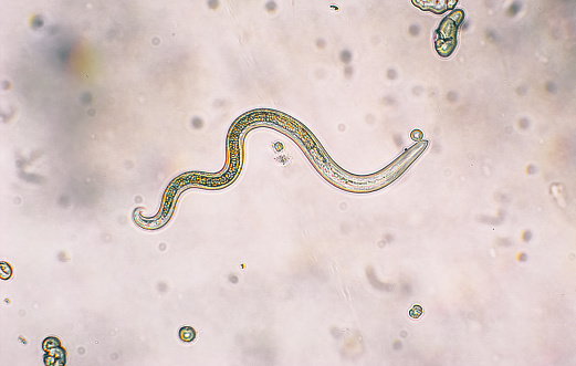
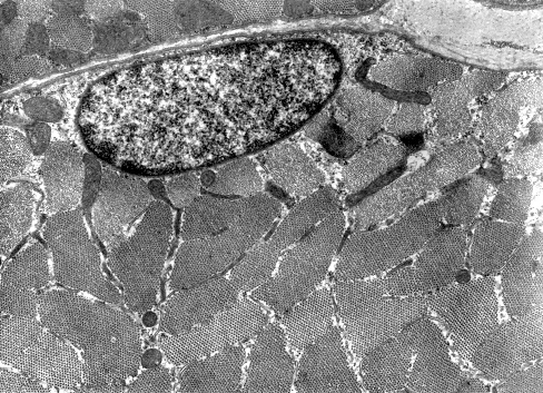
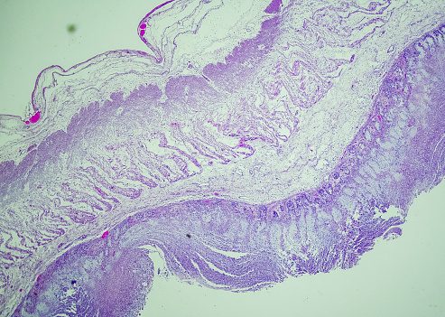
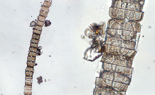
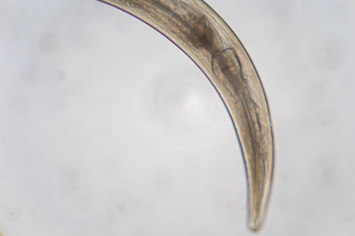
Free Images: "bestof:Schistosoma 20041-300.jpg Electron micrograph of an adult male Schistosoma parasite worm The bar bottom left represents a magnification of 500 μm Photographie"
flea-siphonaptera-insect-parasite-63043.jpg
mite-tuckerella-acari-arachnid-67582.jpg
Schistosoma 20041-300.jpg
Schistosome_Parasite_SEM.jpg
Schistosoma mansoni2.jpg
Schistosoma_mansoni_egg_4841_lores.jpg
Geomyces_destructans.gif
10-0002-F1.png
Terms of Use   Search of the Day