Click Here for More Images from iStock- 15% off with coupon 15FREEIMAGES 
The human eye is an organ that reacts to light and has several purposes. As a conscious sense organ, the mammalian eye allows vision.
Colorful image data that personifies the group
axial lung window chest ct scan 50 years old patient with chest pain showing  bowel  dan gaster visible in  left chest and displacement of the mediastinum to the right
Anatomy scan of twin gestation 6 weeks  monochorionic diamniotic.
Anatomy of human eye. medical concept. 3d illustration
Top view on abstract technical blueprint background with spheres and measurements, wide horizontal composition with copy space
Human eye anatomy. Inner structure. 3d illustration
Volume control on the white background
High contrast shot of woman's eyes and bindi (ornamental dot).
Artificial Intelligence: Simplified image of the principles of convolutional neural networks with input, hidden, output layers. It classifies geometric forms - in this case a cube
Eyes are organs of the visual system. They provide animals with vision, the ability to receive and process visual detail, as well as enabling several photo response functions that are independent of vision.
Doctor holding and showing  human Inner ear model of auditory system
The inner layer of the eye, or retina, is similar to film in a camera. It receives light from an image we are looking at, and converts that light into electrical impulses which are sent through the fibers of the optic nerve to the brain.
Eyes security conversation witness 3D render blue eyeball background.
Eye cancer, eye disease, ophthalmology or optician medical diagnosis concept, 3d illustration
A human anatomy model on a table in a biology classroom for students to study and learn about the structure of the human body.
3D computer graphic sagittal section of female reproductive system with labels.
Soccer ball with national flags on grass over white background. World countries football championship.  3D illustration with copy space. Including clipping path. Set of 26 image
Human eye anatomy on scientific background. 3d illustration
Computational tomography image of the abdominal and pelvic regions.
Bestrophinopathies, inherited retinal disorders caused by mutations in the BEST1 gene, illustration. Best disease, autosomal recessive bestrophinopathy and autosomal dominant vitreoretinochoroidopathy
Mammogram
A PET-CT scan image is a diagnostic visualization combining Positron Emission Tomography (PET) and Computed Tomography (CT) for Helps in finding cancer recurrence.
Printed average efficiency ranking chart  with a magnifying glass .
Eye disease, eye cancer, 3d illustration
Nephrologist holding and examining a human kidney model with stethoscope
Image taken with Flir T420 infra red digital camera. Each color represents different temperatures, as is shown on spectrum scale on right side of image.
3D illustration of a cross-section of the human eye in a side view and a frontal view
Radiology report. On the right, lateral breast there are two intramammary lymph nodes with fatty hilum measuring 0.5 and 0.6 cm respectively. No suspicious breast mass or axillary lymphadenopathy. Moderately dense breasts with 50 to 75 % glandular tissue.  Bi-RADS 2 : benign findings.
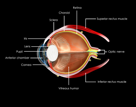
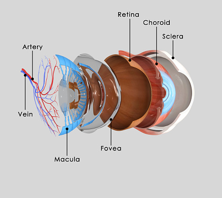
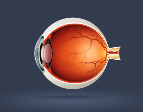
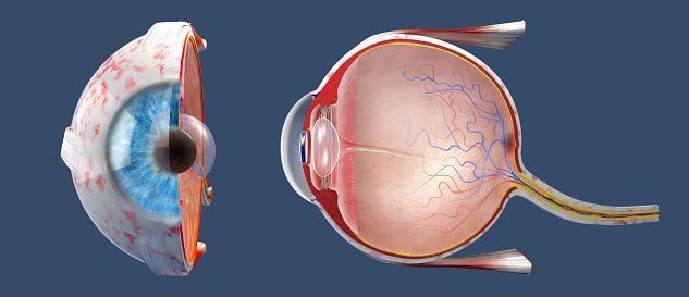
Free Images: "bestof:Schematic diagram of the human eye sk.svg Schematic diagram of the human eye in Slovak Slovak translation of Image Schematic diagram of the human eye en svg by"
Schematic diagram of the human eye sk.svg
Schematic diagram of the human eye el.svg
Schematic diagram of the human eye cs.svg
Schematic diagram of the human eye pl.svg
Schematic diagram of the human eye multilingual.svg
Schematic diagram of the human eye hr.svg
Schematic diagram of the human eye is.svg
Schematic diagram of the human eye ta.svg
Schematic diagram of the human eye en.svg
Schematic diagram of the human eye tr.svg
Schematic diagram of the human eye ar.svg
Schematic diagram of the human eye he.svg
Schematic diagram of the human eye ja.svg
Schematic diagram of the human eye no.svg
Schematic diagram of the human eye it.svg
Schematic diagram of the human eye zh-hans.svg
Schematic diagram of the human eye fr.svg
IOLVIP.svg
Schematic diagram of the human eye pt.svg
Schematic diagram of the human eye horizontal pt.svg
Schematic diagram of the human eye br.svg
Schematic diagram of the human eye cy.svg
Schematic diagram of the human eye pa.svg
Complete neuron cell diagram id.svg
Complete neuron cell diagram fr.svg
Complete neuron cell diagram es.svg
Complete neuron cell diagram de.svg
Complete neuron cell diagram ca.svg
Complete neuron cell diagram en.svg
Complete neuron cell diagram it.svg
Receptive field.svg
Complete neuron cell diagram pl.svg
Complete neuron cell diagram without text.svg
Accommodation (PSF).svg
Complete neuron cell diagram ru.svg
Complete neuron cell diagram numbered large numbers.svg
Feedforward-vs-feedback-driven-eye-movements.svg
Digestive system diagram no labels arrows.svg
Complete neuron cell diagram pt.svg
eye-diagram-eyeball-body-pupil-39998.svg
Complete neuron cell diagram zh.svg
Complete neuron cell diagram zh-hant.svg
Diagram human cell nucleus sk.svg
Schematic diagram of high power fiber laser using a double-clad fiber.svg
Complete neuron cell diagram numbered.svg
Complete diagram of a human spermatozoa pt.svg
Complete diagram of a human spermatozoa pl.svg
Diagram human cell nucleus oc.svg
Deltavs-fr.svg
Fl ru.svg
Human skeleton back en.svg
Human skeleton front en.svg
Human skeleton front sl.svg
Human skeleton back vi.svg
Esquelet humà frontal.svg
Human skeleton front af.svg
Stomach-fr.svg
Digestive system diagram pl.svg
Atmosphaerenzusammensetzung.svg
Human skeleton back pl.svg
Digestive system diagram sk.svg
Circulatory System it.svg
Halftoningcolor.svg
Digestive system diagram lv.svg
Digestive system diagram sr.svg
Digestive system diagram an.svg
Digestive system diagram mk.svg
Digestive system diagram es.svg
Animal mitochondrion diagram id.svg
Gray728.svg
Alveolus diagram.svg
Animal mitochondrion diagram ru.svg
Diagram of the human heart (cropped) bs.svg
Diagram human cell nucleus hu.svg
Human arm bones diagram PL.svg
Diagram human cell nucleus id.svg
Diagram human cell nucleus pt.svg
Diagram human cell nucleus (zh-cn).svg
Diagram human cell nucleus cs.svg
Diagram human cell nucleus tr.svg
Diagram human cell nucleus sl.svg
Competitive inhibitor.svg
ASC schematic.svg
Nanoimprint-en.svg
Pachisi arm schematic.svg
Competitive inhibitor he.svg
Catabolism schematic (zh-cn).svg
Catabolism schematic arm.svg
GasDepositDiagram2.svg
Competitive inhibitor Fi.svg
Mitochondrial electron transport chain pl.svg
Earth-cutaway-schematic-english.svg
Crookes tube2 diagram.svg
Electron transport chain (zh-cn).svg
Digestive system diagram hu.svg
Wiki MagneticBase ALL.svg
Push-pull converter schematic.svg
Silicon purification processes.svg
Circulatory system pl.svg
Principle of the additive mixing.svg
Terms of Use   Search of the Day