Click Here for More Images from iStock- 15% off with coupon 15FREEIMAGES 
Anatomically accurate human male skull with colorized bones lateral or profile view isolated on white background with copy space 3D rendering illustration. Blank anatomical and medical chart.
Panoramic radiography (orthopantomography) by means of X-rays showing the dental structure
Anatomy of the sacrum bone, showcasing its intricate details and features, 3D illustration. Perfect for educational or medical purposes
Axial CT scan through the thorax after intravenous contrast showing the complications which may occur with asbestos exposure. There is extensive left pleural thickening and left pleural fluid due to advanced mesothelioma. Bilateral pleural plaques, some calcified on the left are also seen.
normal head  scan brain window
CAT scan of human abdomen...man with aneurysm of abdominal aorta
Spotted hyena skull, Crocuta crocuta, also known as laughing hyena, is a carnivorous mammal of the family Hyaenidae, of which it is the largest extant member.
CT Scan of an abdomen of a patient with abdominal pain showing swollen appendix suggesting the diagnosis of acute appendicitis.
Canine, Maxillary and Mandibular 3d illustration
Human brain MRI.
An EXCELLENT medical scan taken by a radiologist of a horse's right hoof; imagery was required to make proper diagnosis and determine the extent of the mare's injuries.
Chacma Baboon Skulls at Otjozondjupa Region, Namibia
Side profile view of human skull icon 3d rendering
Panoramic radiography (orthopantomography) by means of X-rays showing the dental structure
The clouded leopard skull on black background.
Plains zebra skull, Equus quagga, formerly Equus burchelli, Common zebra or Burchell's zebra,
Normal chest on computed tomography scan
Close up of a bleached pine marten skull
CAT Scan image pulmonary angiography showing lung adenocarcinoma with red arrow. Medical themes
Computed tomography of chest organs. CT scan. CT Abdomen Cross section View.
Dog skull High quality photo
Skull of a female lion
Lateral Incisor, Maxillar and Mandibular 3d illustration
Computational tomography image of the abdominal and pelvic regions.
Skull of a domestic cat
Human skull of about a 11 to 12 year old showing developing and erupting teeth.
Portrait of an African Lion Skull (Pantera leo).
Skull
Medical imaging
Animal Skulls near Otjiwarongo at Otjozondjupa Region, Namibia
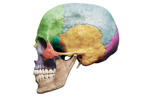
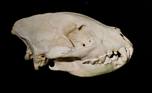
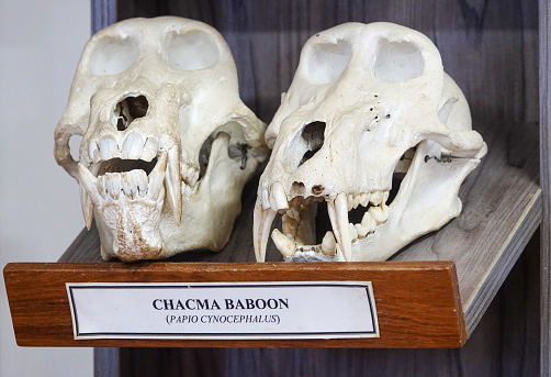
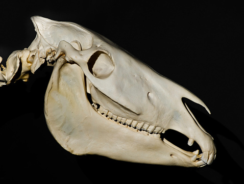
Free Images: "bestof:SKULL-LEFT SIDE - NARA - 524359.jpg Scope and content General notes 524359 Local identifier 106-WB-591 Smithsonian Institution 1846 - NARA-Author O'Sullivan"
Terms of Use   Search of the Day