Click Here for More Images from iStock- 15% off with coupon 15FREEIMAGES 
Epidermis. Electron microscope micrograph showing a keratinocyte of spinous layer. The epithelial cell has a polygonal shape, central nucleus with nucleolus, cytoplasm full of keratin filament bundles, and numerous dark desmosomes crossing the intercellular spaces.
Photomicrograph of algae, Cosmarium turpinii, a desmid. Live specimen. Wet mount, 40X objective, transmitted brightfield illumination.
The study of plant tissues under the microscope in the laboratory.
Blastomycosis is a fungal infection caused by inhaling Blastomyces dermatitidis spores. If it involves only the lungs, it is called pulmonary blastomycosis. Only about half of people with the disease have symptoms, which can include fever, cough, night sweats, muscle pains, weight loss, chest pain, and feeling tired.
Thyroid cancer: Microscopic image of Metastatic papillary carcinoma of thyroid. Reactive change. Lymph node carcinoma.
Kidney proximal tubule epithelial cloudy SWELLING under different area in light microscopyKidney
PAPS Smear study of a young women under microscopy showing Atrophic changes in uterus which is unusual at young age.
Monkeypox Virus. 3D Render
geranium leaf under microscope showing trichomes - optical microscope x200 magnification
Quantum Gravity 3d rendered image of a comprehensive exploration of Quantum Gravity theories. String theory, loop quantum gravity (LOG), causal dynamical triangulation, twistor theory, gravitational waves, spacetime, gravity waves, quantum mechanics, quantum entanglement concepts.
Leiden leaf c.s. under microscope
Myxoid liposarcoma.  Myxoid/round cell liposarcoma, or MRCLS, is one of several types of liposarcoma. Liposarcoma is a rare cancer that grows in the cells that store fat in the body. MRCLS usually grows in the arms and legs. These tumors grow slowly, and they can spread to other parts of the body
Pine mature wood cross section. Light microscope slide with microsection of an evergreen conifer in the genus Pinus. Plant anatomy. Biology. Photo.
Natural agate surfaces, suitable for backgrounds and wallpapers.
Prostate Cancer: Microscopic image of prostatic tissue, adenocarcinoma, malignant neoplasm, atypical epithelial cells.
Close up of cristal beads
Backgrounds of Bacteria and Mold under the microscope in Lab.
Microscopic photo of a thin section of calcareous tufa of Holocene age.
Squash preparation of  onion root tip cells stained to reveal chromosomes,  Bright field illumination, light microscopy.  This image was taken with the X40 objective lens.  Depth of focus is extremely low at this magnification.  Smaller apertures give greater depth of focus, but image quality deteriorates and resolution is greatly reduced.  The aperture in this image was optimised for contrast and resolution. Chromatic abberation is inevitable in light microscopy, but has been kept to a minimum in this image.
Metastatic plasmacytoma, metastatic undifferentiated sarcoma. Microscopically show cellular material of mostly plasmacytiod cells. atypical mitoses are present. plasma cell tumor.
Hepatocellular carcinoma, HCC under microscopy
Plants cell texture.
corn stem micrograph with dye
Histological Pancreas human, Liver human, Vermiform appendix human and Kidney Human under the microscope for education.
Focal nodular hyperplasia (FNH) is a benign liver lesion that is composed of a proliferation of hyperplastic hepatocytes surrounding a central stellate scar. Typically, FNH is a solitary lesion that is more commonly seen in women.
Diatoms, algae under microscopic view, phytoplankton, fossils, silica, golden yellow algae
Particles, medical concept
Breast tissue adenocarcinoma immunohistochemistry. Estrogen receptors.
plant Zea Stem C.S. under light mircoscope with white background
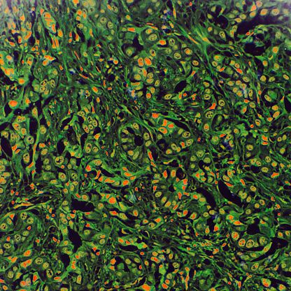
Free Images: "bestof:S cerevisiae septins.jpg Fluorescent micrograph of Saccharomyces cerevisiae with GFP-tagged septins <br /> Green AgSEP7-GFP septin red outline of the cell phase"
S cerevisiae septins.jpg
A gossypii septins.jpg
A gossypii.jpg
Ascus.gif
Shmoo yeast S cerevisiae.jpg
Alpha-Amanitin–RNA polymerase II complex 1K83.png
HeLa pspecks2.jpg
Phase contrast image of L-form bacteria-Mark Leaver Newcastle University.tif
Fluorescent image fibroblast.jpg
Organotypic chicken spinal cord.tiff
Mouse NT antibody NF Ki67.jpg
Transmission line animation.gif
Terms of Use   Search of the Day