Click Here for More Images from iStock- 15% off with coupon 15FREEIMAGES 
Leiden leaf c.s. under microscope
Colorful image data that personifies the group
A 3D medical illustration of the sacrum bone isolated on a white background, showcasing its shape, structure, and anatomical features. Back view
Carcinoid tumors are a type of slow-growing cancer that can arise in several places throughout your body. Carcinoid tumors, which are one subset of tumors called neuroendocrine tumors, usually begin in the digestive tract (stomach, appendix, small intestine, colon, rectum) or in the lungs. Site: Ovary
The vertebral column surrounds the spinal cord which travels within the spinal canal, formed from a central hole within each vertebra
Glutenous Maximums: This is the largest and most superficial of the gluten muscles. It is responsible for the extension and lateral rotation of the hip joint. The glutenous maximums is what gives the buttocks its shape and provides power during activities like running, climbing, and jumping.
The hud interface of the futuristic medical program is a control panel with digital three-dimensional MRI visualization, which allows you to study the condition of joints, spine, muscles and soft tissues in detail.
The study of plant tissues under the microscope in the laboratory.
Doctor holding and showing  human Inner ear model of auditory system
Human anatomy. Thyroid gland in human body on white. Thyroid control.
Metastatic neuroendocrine tumor. Site: Liver.  Neuroendocrine tumors are cancers that begin in specialized cells called neuroendocrine cells. Neuroendocrine cells have traits similar to those of nerve cells and hormone-producing cells. Neuroendocrine tumors are rare and can occur anywhere in the body.
3D illustration of  human body shape (female). Useful for art, health-care, product design etc.
The detrusor muscle remains relaxed to allow the bladder to store urine, and contracts during urination to release urine 3d illustration
Digitally generated image of a human hologram walking on a dark background with a coop space for a futuristic scientific design
Cross-section Dicot, Monocot and Root of Plant Stem under the microscope for classroom education.
skin science 3d
Urology, treatment of men diseases and prostatitis. Consultation of a male urologist for a patient with prostatitis
corn stem micrograph with dye
The torso or trunk is an anatomical term for the central part, or the core, of the body of many animals, from which the head, neck, limbs, tail and other appendages extend.
a visual aid of the inside of the human head isolated on white.
The fifth lumbar spine vertebrae (L5) is part of the greater lumbar region 3d illustration
Abstract background of cell
PAPS Smear study of a young women under microscopy showing Atrophic changes in uterus which is unusual at young age.
Squamous cell carcinoma of the lung is a type of lung cancer. It occurs when abnormal lung cells multiply out of control and form a tumor.
3D Illustration of Human Skeleton System Pelvis with Labels Anatomy (Posterior View)
Medical concept
Human Skeleton Hip or Pelvic bone Anatomy For Medical Concept 3D Illustration
3D representation of the human body
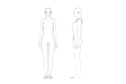
Free Images: "bestof:Risting keja.jpg en Human male and female - anatomical features pointed out Human_anatomy jpg 2010-01-24 23 36 UTC Human_anatomy jpg User Ralf Roletschek / User"
Terms of Use   Search of the Day