Click Here for More Images from iStock- 15% off with coupon 15FREEIMAGES 
3D Illustration of Female Reproductive System
Cropped image of screen showing researcher looking in a microscope fertilising an egg. Intracytoplasmic sperm injection.
Polycystic Ovarian Syndrome or PCOS on ovaries symptoms as hormonal disorder with small ovarian cysts as a concept of female fertility or infertility with fallopian tubes.
Anatomy scan of twin gestation 6 weeks  monochorionic diamniotic.
Doctor holding human Colon anatomy model. Colonic disease, Large Intestine, Colorectal cancer, Ulcerative colitis, Diverticulitis, Irritable bowel syndrome, Digestive system and Health concept
2D pregnancy ultrasound of a baby at 30 weeks
Kidney 3D Icon. Human kidney internal organ 3d illustration
skin science 3d
Bacterial vaginosis is a genital infection caused by bacteria, mainly Gardnerella Vaginalis. 3D illustration
Gynecologist holding and showing an ovary model
Human bladder cancer as a urinary anatomical organ disease and malignant cells concept as a 3D illustration cutaway of body anatomy.
Liver Fluke Section under light microscope with white background
Full size digital screening mammogram of a new breast tumor in the retroareolar region of the right breast causing nipple retraction. An ultrasound guided biopsy was performed diagnosing invasive ductal carcinoma. This is the most common type of breast cancer, accounting for nearly 80% of cases. Breast cancer treatment has come a long way in the last few decades, seeing more funding than any other human disease. The first step to a cure is catching it early with screening mammograms such as this case so that the tumor can be removed before it spreads (metastasizes) out of the breast.
3D Illustration Concept of Human Urinary System Bladder Anatomy
Prostate and male reproductive system isolated on white
Medical concept
Hologram of the female organ of the uterus on a blue background. Ultrasound concept, gynecology, obstetrics, ovulation, pregnancy. 3D illustration, 3D render
Plasmacytoid urothelial carcinoma (PUC) is a unique variant of invasive urothelial carcinoma characterized by tumor cells that exhibit a striking resemblance to plasma cells, and it is known to have an aggressive behavior.
Acid Reflux GERD Disease Illustration
7-month fetus in womb
prostate cancer, prostatic hyperplasia, Structure of male prostate, cross section, 3d illustration
Acute ordinary hepatitis liver biopsy under light microscopy
axial lung window chest ct scan 50 years old patient with chest pain showing  bowel  dan gaster visible in  left chest and displacement of the mediastinum to the right
Four, side view, X-ray pictures of a woman's two breasts. On the right, lateral breast there are two intramammary lymph nodes with fatty hilum measuring 0.5 and 0.6 cm respectively. No suspicious breast mass or auxiliary lymphadenopathy. Moderately dense breasts with 50 to 75 % glandular tissue.  Bi-RADS 2 : benign findings.
Doctor holding human Colon anatomy model. Colonic disease, Large Intestine, Colorectal cancer, Ulcerative colitis, Diverticulitis, Irritable bowel syndrome, Digestive system and Health concept
Gynecologist holding and examining an ovary model symbolizing annual check up
Ovaries concept and uterus with fallopian tubes as a magnification close up of a human female reproduction illustration on a white background as a symbol of fertility and reproductive system health.
Pheochromocytoma, Site: Adrenal gland
Uterus: A hollow, pear-shaped organ that is located in a woman's lower abdomen, between the bladder and the rectum. The narrow lower portion of the uterus is the cervix (the neck of the uterus). The broader upper part is the corpus, which is made up of three layers of tissue.
Macro close up depicting the abstract forms and shapes in a mixture of grey paint and oil.
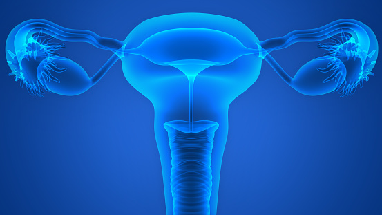
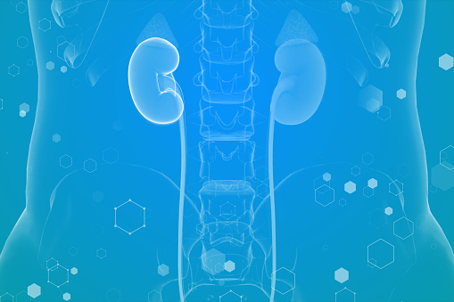
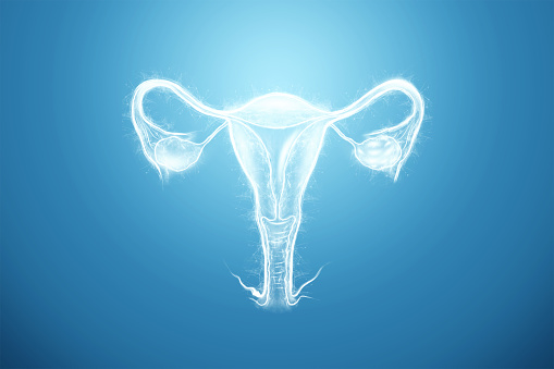
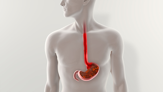
Free Images: "bestof:RLQ2unlabled.PNG image of RLQ containing Cecum Appendix Right ovary and tube Right ureter work File Digestive_system_diagram_en svg by Mariana Ruiz Villarreal"
Sadf.svg
Human skull side bones tr.svg
Esquelet humà frontal.svg
Scheme human hand bones-en.svg
Right Ventricular hypertrophy cs.svg
PD Diagram of swine flu symptoms EN.svg
Diagram of influenza infection symptoms EN.svg
Diagram of swine influenza symptoms EN.svg
Human skull side suturas right pl.svg
Human skull side suturas right heb.svg
Human skull front bones numbered.svg
Human skull side details right pl.svg
Human skull side suturas pl.svg
Human skull side details.svg
Human skull side details pl.svg
Human skull side complete.svg
Metacarpals numbered-en (left hand).svg
Os du crane.svg
Metacarpals numbered-en.svg
Human skull side complete pl.svg
Average prokaryote cell- ru.svg
Average prokaryote cell- uk.svg
Diagram human cell nucleus oc.svg
Diagram human cell nucleus id.svg
Entamoeba histolytica life cycle-pl.svg
Circulatory System it.svg
Scheme human hand bones-ru.svg
Scheme human hand bones-en-ru.svg
Circulatory System en edited.svg
Skull template.svg
Human skeleton front sl.svg
Human skeleton back sl.svg
Human skull side suturas.svg
Human skull side details right.svg
Plastids types en.svg
Complete diagram of a human spermatozoa pt.svg
Phospholipide Structuren ger.svg
Animal mitochondrion diagram id.svg
Scheme sodium-potassium pump-it.svg
Scheme sodium-potassium pump-ca.svg
Human skeleton back en.svg
Human skeleton front ru.svg
Average prokaryote cell- id.svg
Leishmaniasis life cycle diagram ar.svg
Circulatory System ru.svg
Human skeleton front en.svg
Appendicular skeleton diagram blank.svg
Scheme sodium-potassium pump-en-2.svg
Scheme sodium-potassium pump-zh.svg
Human skeleton back ru.svg
Culex mosquito life cycle uk.svg
Phospholipids aqueous solution structures ES.svg
Culex mosquito life cycle es.svg
Leishmaniasis life cycle diagram ru.svg
Scheme sodium-potassium pump-de.svg
Animal mitochondrion diagram zh hk.svg
Animal mitochondrion diagram zh tw.svg
Scheme sodium-potassium pump-he.svg
Babesia life cycle human de.svg
Human skull side simplified (bones) ku.svg
Culex pipiens diagram uk.svg
Human skull side simplified (bones) ru.svg
Human skull front simplified (bones) ru.svg
Scheme simple diffusion in cell membrane-zh (zh-cn).svg
Scheme facilitated diffusion in cell membrane-zh.svg
Animal mitochondrion diagram ml.svg
Animal mitochondrion diagram zh ml.svg
Scheme facilitated diffusion in cell membrane-ca.svg
Human skull side suturas right.svg
Scheme simple diffusion in cell membrane-pl.svg
Animal mitochondrion diagram ru.svg
Culex pipiens diagram fr.svg
Leishmaniasis life cycle diagram-es.svg
Scheme simple diffusion in cell membrane-ca.svg
Entamoeba histolytica life cycle-it.svg
Axial skeleton diagram blank.svg
Scheme secundary active transport-heb.svg
Pectoral girdle front diagram de.svg
Scheme secundary active transport-pl.svg
Strutture fosfolipidiche in soluzione acquosa.svg
Culex pipiens diagram es.svg
Pectoral girdle front diagram nan.svg
Human skeleton front arrows no labels.svg
Prokaryote cell-ro.svg
Scheme simple diffusion in cell membrane-zh (zh-tw).svg
Average prokaryote cell- nl.svg
Scheme sodium-potassium pump-cs.svg
Right Ventricular hypertrophy-IT.svg
Endocytosis types-fr.svg
Human skull front bones ku.svg
Human skull nolables.svg
Scheme sodium-potassium pump-pl.svg
Mature flower sv blue-labels.svg
Tree of life SVG.svg
Wine grape diagram en.svg
Plastids types uk.svg
Cell junction simplified zh traditional.svg
Pinocytosis gl.svg
Mature flower numbered.svg
Plant cell structure svg zh-hant.svg
Terms of Use   Search of the Day