Click Here for More Images from iStock- 15% off with coupon 15FREEIMAGES 
3d rendered medically accurate illustration of a female brain anatomy
An acupuncture model of the male human torso depicting the acupoints and meridiens used in Traditional Chinese Medicine and massage therapy
Dopamine is a chemical messenger and neurotransmitter produced in the brain that affects many bodily functions. It's an organic chemical that's part of the catecholamine and phenethylamine families. Dopamine is responsible for many effects, including mood, movement, memory, motivation, attention.
Medical illustration series about abstract human.The nervous system.
The central nervous system is the brain and spinal cord, while the peripheral nervous system consists of everything else 3d illustration
Cyanobacteria or blue-green algae,
In biology, the classical doctrine of the nervous system determines that it is a highly complex part of an animal that coordinates its actions and sensory information by transmitting signals to and from different parts of its body. The nervous system detects environmental changes that impact the body, then works in tandem with the endocrine system to respond to such events.
Invasive pleomorphic lobular carcinoma (PLC) of the breast is a subtype of invasive lobular cancer which compromises approximately 1% of all epithelial breast malignancies and is characterized by higher nuclear pleomorphism and poorer prognosis than classic invasive lobular cancer (ILC)
Human skull showing nerves, tongue and part of the cerebellum. Medicine illustration
Section of epididymis under light microscope with white background
3D Illustration Human Brain Anatomy With Nervous System For Medical Concept
Doctor holding a  human head model
The external spermatic fascia (Intercrural Fascia intercolumnar fascia) is a thin membrane,
Digital medical illustration: Posterior (back) view (orthogonal) of human skull (includes teeth) and neck featuring vertebrae, discs and ligaments (includes visible Interspinale, Nuchae, Anulus, Flavum, Capsula and Intertransversarium).
3d rendered medically accurate illustration of male head  nervous system and skeleton system
Carcinoid tumors are a type of slow-growing cancer that can arise in several places throughout your body. Carcinoid tumors, which are one subset of tumors called neuroendocrine tumors, usually begin in the digestive tract (stomach, appendix, small intestine, colon, rectum) or in the lungs. Site: Ovary
Gut-brain connection or gut brain axis. Concept art showing a connection from the gut to the brain. 3d illustration.
Firing Neurons - 3d rendered image of Neuron cell network on black background.  Conceptual medical illustration.  Healthcare concept. \nAbstract stylized npr (non photorealistic rendering) view. Half Tone, Spotted, Stipple Effect. Funky, popart, retro style colors.
Showing Light micrograph Type of Tissue Human under the microscope in Lab.
Magnetic resonance images of the brain (MRI brain). Computed resonance imaging of the human brain.
The central nervous system is the brain and spinal cord 3d illustration
a visual aid of the human brain isolated on white.
The central nervous system is made up of the brain and spinal cord 3D illustration
Adenocarcinoma, predominantly papillary type, lung
A brain is an organ that serves as the center of the nervous system in all vertebrate and most invertebrate animals. It is located in the head, usually close to the sensory organs for senses such as vision. It is the most complex organ in a vertebrate's body.
plant Pine Cone-Female L.S. under light mircoscope with white background
Cross section of vertebrae. Medical background. 3d illustration
X-ray image of a human brain made up mostly of water
Computer Generated Image: Sideview of a transparent head of a man with the brain parts isolated and labeled against a white background.
skin science 3d
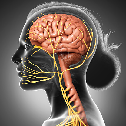
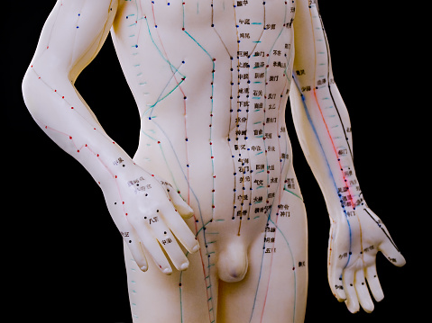
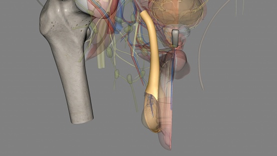
Free Images: "bestof:Pudendal nerve.svg Pudendal nerve course and branches Essential Clinical Anatomy K L Moore A M Agur Lippincott 2 ed 2002 Page 263 2008-01-26 Mikael Häggström"
Pudendal nerve.svg
Pudendal nerve-es.svg
Stomach blood supply.svg
Es-Stomach blood supply.svg
Glycogen structure.svg
Notable mutations.svg
Health effects of cheese.svg
Effects of sleep deprivation.svg
road-branch-aesthetic-nature-kahl-946490.jpg
Gray812and814.svg
Maxillary_artery_branches.svg
Gray812and814 - PT.svg
Amino acid catabolism revised.svg
Lymphocyte activation.svg
Surface projections of the organs of the trunk - without labels.svg
Female shadow anatomy without labels.svg
Male template with organs.svg
Female template with organs.svg
Surface projections of the organs of the trunk.svg
Dermatomes and cutaneous nerves - posterior.svg
Renin-angiotensin system in man shadow (zh-cn).svg
Mandibular_nerve_branches.png
Glycogen structure.png
Gray819.png
Greater_splanchnic_nerve.png
Health effects of cheese.png
Image_from_page_554_of_"Local_and_regional_anesthesia_:_with_chapters_on_spinal,_epidural,_paravertebral,_and_parasacral_analgesia,_and_on_other_applications_of_local_and_regional_anesthesia_to_the_surgery_of_the_eye,_ear,_nose_and_throat,_and_to_dental_p.jpg
Effects of sleep deprivation.png
Anatomy of Ulnar nerve.JPG
VIIth Nerve.jpg
Gray794.png
Gray794A.png
Gray789.png
Gray779.png
Mandibular branches.PNG
Infraorbital_artery.png
Masseteric_artery.png
Illu_diencephalon.jpg
Turbellaria_001.png
Ascending_and_descending_branches.PNG
Great_saphenous_vein.png
Maxillary_artery_branches.png
Anterior_tympanic_artery.png
Buccal_artery.png
Deep_temporal_arteries.png
Inferior_alveolar_artery.png
Middle_meningeal_artery.png
Posterior_superior_alveolar_artery.png
Pterygoid_branches_of_maxillary_artery.png
Axillary_branches.PNG
Internal_iliac_branches.PNG
Superficial_and_deep_branches.png
Gray812and814.PNG
Gray812and814_hand.png
Axillary_branches_es.png
Lymphocyte activation.png
Amino acid catabolism revised.png
Flatau_atlas1984_en.jpg
Limites_artère_axillaire.png
External_maxillary_artery_branches.png
Inferior_thyroid_and_ascending_cervical.PNG
Female shadow anatomy without labels.png
Gray761.png
Gray926.png
Lymphocyte activation-es.png
Ascidia mentula 002.png
Fundus photograph of normal left eye.jpg
Subclavius_1st_part.png
Costocervical_trunk_with_branches.png
FMIB_47895_Left_crusher_claw_of_lobster,_partly_dissected_from_upper_side,_to_show_relations_of_muscles,_nerves,_blood_vessels,_and_skin.jpeg
Fundus photograph of normal right eye.jpg
Dermatomes and cutaneous nerves - posterior.png
Anatomy_of_an_earthworm_001.png
Handel,_Rinaldo_Aria,_1876.png
Doliolum_denticulatum_001.png
Dr. Mosstaghimi.jpg
Annelid Cross-sect 02.png
Rhopalium in Selected morphological monographs - The Cubomedusae (1900).png
Surface projections of the organs of the trunk.png
A-granulatum (Alvesgaspar) B epimera.jpg
A-granulatum (Alvesgaspar) A flagellum.jpg
A-granulatum (Alvesgaspar) A flagellum zoom.jpg
A-granulatum (Alvesgaspar) B flagellum.jpg
A-granulatum (Alvesgaspar) B flagellum zoom.jpg
A-granulatum (Alvesgaspar) C flagellum.jpg
A-granulatum (Alvesgaspar) C flagellum zoom.jpg
A-granulatum (Alvesgaspar) A head front.jpg
A-granulatum (Alvesgaspar) A telson.jpg
A-granulatum (Alvesgaspar) D head top.jpg
ChronicBackache1040.png
Beckert's_garden_field_and_flower_seeds_(16373608768).jpg
ChronicBackache1041.png
Everything_for_the_garden_(16205078337).jpg
Terms of Use   Search of the Day