Click Here for More Images from iStock- 15% off with coupon 15FREEIMAGES 
Green algae cells background
Trematode parasite under a microscope
The Raf/MEK/ERK (MAPK) propagates growth signals in the cell. Misregulation can lead to cancer.\nThe red dots on the active EGFR are phosphorylated tyrosines. PDB entries: 1egf, 1nql, 1m17, 2jwa, 3njp, 2gs6, 1gri, 1xd2, 3ksy, 5p21, 6xi7, 6q0j, 2y4i,
Micrograph of amoeba.  Live specimen. Wet mount, 40X objective, transmitted brightfield illumination. Note - motion blur of live animal, very shallow depth of field, chromatic aberration and uneven focus are inherent in light microscopy.
Bacteria Aggregatibacter, 3D illustration. Aggregatibacter aphrophilus and A. actinomycetemcomitans, Gram-negative bacteria, part of the normal flora of the mouth and throat, also cause endocarditis
Squash preparation of stained onion root tip cells showing various stages of mitosis. Differential interference contrast optics on a Leica microscope, Image taken with the X40 objective lens.  Depth of focus is extremely shallow (just a few microns).  Differential interference contrast results in an optical slice horizontally through the specimen providing maximum possible resolution at this magnification and good contrast. Chromatic abberation is inevitable in light microscopy, but has been kept to a minimum in this image.
Diatoms, algae under microscopic view, phytoplankton, fossils, silica, golden yellow algae
abs Bacteria concept - 3d rendered microbiology image. Medical research, health-care concept.
Host cells with spores (mold) are inside wood under the microscope for education.
Myxoid liposarcoma.  Myxoid/round cell liposarcoma, or MRCLS, is one of several types of liposarcoma. Liposarcoma is a rare cancer that grows in the cells that store fat in the body. MRCLS usually grows in the arms and legs. These tumors grow slowly, and they can spread to other parts of the body
After dopamine binding, the dopamine receptor changes his shape and binds the inactive G protein.The G protein expels its GDP molecule replacing it by GTP. and falls into two pieces. The Galpha subunit binds to and activates the enzyme adenylyl cyclase, which produces then cyclic AMP spreading a signal through the cell.
Liver Fluke Section under light microscope with white background
Escherichia Coli (E. Coli.) bacteria under microscope
Close-up shot of a petri dish for checking an antibiotic in the laboratory.
Leiden leaf c.s. under microscope
Bacteria pathogen - 3d rendered microbiology image. Illustrate of Lactobacillus Bulgaricus Bacteria, Microbacterium Tuberculosis Bacteria (MTB). Medical research, health-care concept. SEM (TEM) scanning view
Photomicrograph of algae, Cosmarium turpinii, a desmid. Live specimen. Wet mount, 40X objective, transmitted brightfield illumination.
Freshwater aquatic zooplankton and algae under microscope view
The intermembrane transporter of the Gram-negative bacterium Acinetobacter baylyi transports a lipopolysaccharide (orange) through the inner membrane consuming ATP (red)
Green chlorophyll Cells under heavy magnification isolated on dark background
Freshwater Closterium algae (unicellular charophyte green algae) - optical microscope x200 magnification
Squash preparation of stained onion root tip cells showing various stages of mitosis. Differential interference contrast optics on a Leica microscope, Image taken with the X40 objective lens.  Depth of focus is extremely shallow (just a few microns).  Differential interference contrast results in an optical slice horizontally through the specimen providing maximum possible resolution at this magnification and good contrast. Chromatic abberation is inevitable in light microscopy, but has been kept to a minimum in this image.
Hand fungal infection. Ringworm, tinea manuum and close-up view of dermatophyte fungi Microsporum canis, Microsporum audouinii, Trichophyton rubrum and Trichophyton mentagrophytes, 3D illustration
Abs Bubonic plague bacteria - 3d rendered image structure view on black background. \nPlague Infection concept.  Hologram bacteria view.
Eggs of helminth and Finding parasites in feces, analyze by microscope in laboratory.
In vitro bacterial culture
Fluconazole is an antifungal medication used to treat fungal infections like candidiasis and mycosis. Fluconazole alters the active site of the lanosterol 14 alpha-demethylase preventing the demethylation of lanosterol, an essential step in the biosynthesis of ergosterol. Ergosterol plays an important structural role in the regulation of membrane fluidity and permeability. Source: PDB entry 5esf.
The pale lactose nonfermenting bacterie (blue arrow) are Salminella typhi.  Salmonella in general does not ferment lactose (colorless colonies),  Salmonella typhi,unlike other salmonallae, produce little hydrogen sulfide (H2S) gas.  The resulting bacterial colonies will appear colorless with colorless or very discrete black centers.
Photomicrograph of filamentous blue-green algae, Oscillatoria species. Each band along the stand is one cell. This filament is very short probably having recently parted from a longer one. Filaments are typically many times longer. This algae is mobile, it moves and twists through the water. San Francisco Bay, California, USA. Live specimen. Wet mount, 40X objective, transmitted brightfield illumination. Note - motion blur of live specimen, very shallow depth of field, chromatic aberration and uneven focus are inherent in light microscopy.
Microalgae under microscopic view, green algae, cyanobacteria, phytoplankton, diatom, algae mix collage background
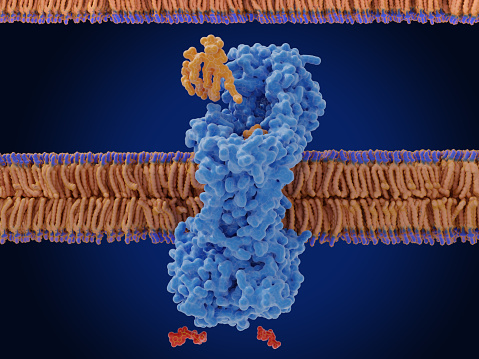
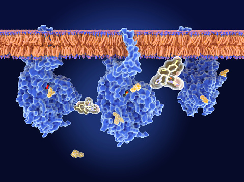
Free Images: "bestof:Porcine Mitochondrial IDH R stabilization.jpg Isocitrate Dehydrogenase enzyme active site and Arg AA stabilization of Isocitrate From PDB 1LWD Created using"
P450cycle.svg
Bondarzewiaceae Maximum Likelihood-Tree .svg
Porcine Mitochondrial IDH R stabilization.jpg
Porcine Mitochondrial IDH surface with greenpocket.jpg
Porcine Mitochondrial IDH surface pocket view.jpg
Porcine Mitochondrial IDH ball view.jpg
Porcine Mitochondrial IDH active site waters.jpg
Porcine Mitochondrial IDH H donation to O3.jpg
Porcine Mitochondrial IDH mn interaction.jpg
Ecoli IDH with surface pocket.jpg
Porcine Mitochondrial IDH 1lwd.jpg
Isocitrate_dehydrogenase_(coli).jpg
IDHcatalyticmechanism.jpg
Isocitrate dehydrogenase of Escherichia coli Complex.jpg
Citrate synthase 1csi.png
Isocitrate dehydrogenase2.png
Isocitrate dehydrogenase.png
PDB 1lwd EBI.jpg
IsocitratedehydrogenaseloseH.gif
Isocitratedehydrogenaseakg.gif
Isocitratedehydrogenaselosedb.gif
Isocitrate lyase.png
Succinate Dehydrogenase.jpg
Aconitase center 7ACN.png
Carbonic anhydrase 1CA2.png
Carbonic anhydrase 1CA2 active site.png
Alpha-Amanitin–RNA polymerase II complex 1K83.png
Ketoconazole 3D balls 1jin.png
Isocitrate dehydrogenase 1ai3.png
Malate Dehydrogenase Active Site.jpg
Cys and Arg active site of ubiquitin activating enzyme.png
PDB 1xab EBI.jpg
PDB 1xaa EBI.jpg
PDB 1idm EBI.jpg
PDB 1g2u EBI.jpg
PDB 1gc9 EBI.jpg
PDB 1osj EBI.jpg
PDB 1wal EBI.jpg
PDB 1ipd EBI.jpg
PDB 1gc8 EBI.jpg
PDB 1osi EBI.jpg
PDB 1dr0 EBI.jpg
PDB 1dr8 EBI.jpg
PDB 1dpz EBI.jpg
PDB 1hex EBI.jpg
PDB 1xad EBI.jpg
PDB 1xac EBI.jpg
PDB 2ayq EBI.jpg
PDB 1v53 EBI.jpg
PDB 1a05 EBI.jpg
PDB 1cnz EBI.jpg
PDB 1cm7 EBI.jpg
PDB 1vlc EBI.jpg
PDB 1wpw EBI.jpg
PDB 1w0d EBI.jpg
PDB 2g4o EBI.jpg
PDB 1pb1 EBI.jpg
PDB 1pb3 EBI.jpg
PDB 1iso EBI.jpg
PDB 1p8f EBI.jpg
PDB 1cw1 EBI.jpg
PDB 1cw4 EBI.jpg
PDB 1sjs EBI.jpg
PDB 1cw7 EBI.jpg
PDB 7icd EBI.jpg
PDB 1ai2 EBI.jpg
PDB 1ai3 EBI.jpg
PDB 1idc EBI.jpg
PDB 9icd EBI.jpg
PDB 8icd EBI.jpg
PDB 4icd EBI.jpg
PDB 1gro EBI.jpg
PDB 3icd EBI.jpg
PDB 1grp EBI.jpg
PDB 5icd EBI.jpg
PDB 6icd EBI.jpg
PDB 1hj6 EBI.jpg
PDB 1bl5 EBI.jpg
PDB 1idd EBI.jpg
PDB 1idf EBI.jpg
PDB 1ika EBI.jpg
PDB 1ide EBI.jpg
PDB 1hqs EBI.jpg
PDB 1j1w EBI.jpg
PDB 1t0l EBI.jpg
PDB 1t09 EBI.jpg
PDB 1ptm EBI.jpg
PDB 1ps6 EBI.jpg
PDB 1ps7 EBI.jpg
PDB 1r8k EBI.jpg
PDB 1vi1 EBI.jpg
PDB 1u7n EBI.jpg
PDB 2af4 EBI.jpg
PDB 1qzt EBI.jpg
PDB 2af3 EBI.jpg
PDB 1r5j EBI.jpg
PDB 1xco EBI.jpg
PDB 1td9 EBI.jpg
PDB 1vmi EBI.jpg
PDB 1itw EBI.jpg
Terms of Use   Search of the Day