Click Here for More Images from iStock- 15% off with coupon 15FREEIMAGES 
Nirmatrelvir inhibits the 3C-like protease of the SARS-CoV 2 virus. The coronavirus 3CL hydrolase enzyme, also known as the main protease, is essential for the maturation of the virus.
The three-dimensional structure of proteins is represented in a variety of conformations. This image is based on the ribbon structure of a protein. It is suitable for biology notebooks and book covers.\nThis image has not been created by AI.
Bacteria Aggregatibacter, 3D illustration. Aggregatibacter aphrophilus and A. actinomycetemcomitans, Gram-negative bacteria, part of the normal flora of the mouth and throat, also cause endocarditis
Escherichia Coli (E. Coli.)
3d helix chromosome or Dna structure, technology science background. 3d render illustration
Photomicrograph of filamentous blue-green algae, Oscillatoria species. Each band along the stand is one cell. This filament is very short probably having recently parted from a longer one. Filaments are typically many times longer. This algae is mobile, it moves and twists through the water. San Francisco Bay, California, USA. Live specimen. Wet mount, 40X objective, transmitted brightfield illumination. Note - motion blur of live specimen, very shallow depth of field, chromatic aberration and uneven focus are inherent in light microscopy.
Dna structure on science background. 3d illustration
Abs Bubonic plague bacteria - 3d rendered image structure view on black background. \nPlague Infection concept.  Hologram bacteria view.
Bacterial multidrug efflux pumps (E-CmeB) expel antibiotics  from inside the bacterium (bottom): ciprofloxacin (yellow), chloramphenicol (violet),  ampicillin (red).
DNA, Adenine, Cytosine, Guanine, Laboratory
Cell membrane and biology, biological concept, 3d rendering. Computer digital drawing.
In vitro bacterial culture
B-chain pentamer and A-chain subunit (green), 3D cartoon and Gaussian surface model, white background
Close up of cristal beads
Escherichia Coli (E. Coli.) bacteria under microscope
Endosomes - 3d rendered image of Endosomes are membrane-bound organelles. They are parts of endocytic membrane transport pathway.  Endomembrane system in cells. Bilayer Lipid Membrane. Abstract medical illustration. Microscopic view. Microbiologic research concept.
Green algae cells background
Liver Fluke Section under light microscope with white background
Licmophora sp. algae, marine and freshwater diatom under microscopic view. Genus of benthic, photosynthetic and epiphyte diatom
Chain or ribbon diatom, a type of algae.
Backgrounds of Bacteria and Mold under the microscope in Lab.
Squash preparation of stained onion root tip cells showing various stages of mitosis. Differential interference contrast optics on a Leica microscope, Image taken with the X40 objective lens.  Depth of focus is extremely shallow (just a few microns).  Differential interference contrast results in an optical slice horizontally through the specimen providing maximum possible resolution at this magnification and good contrast. Chromatic abberation is inevitable in light microscopy, but has been kept to a minimum in this image.
Freshwater Closterium algae (unicellular charophyte green algae) - optical microscope x200 magnification
Monkeypox Virus. 3D Render
A quadrant streak of the bacterium Staphylococcus aureus cultured or inoculated on Baird-Parker agar with egg tellurite .
Bacteria pathogen - 3d rendered microbiology image. Illustrate of Lactobacillus Bulgaricus Bacteria, Microbacterium Tuberculosis Bacteria (MTB). Medical research, health-care concept. SEM (TEM) scanning view
Colorful chain of amino acids or bio molecules called proteins - 3d illustration
Myxoid liposarcoma.  Myxoid/round cell liposarcoma, or MRCLS, is one of several types of liposarcoma. Liposarcoma is a rare cancer that grows in the cells that store fat in the body. MRCLS usually grows in the arms and legs. These tumors grow slowly, and they can spread to other parts of the body
3D cartoon and Gaussian surface models, PDB 6gu2, white background
plant Flower bud of Lilium brownii var.viridulum T.S.\nunder light mircoscope with white background
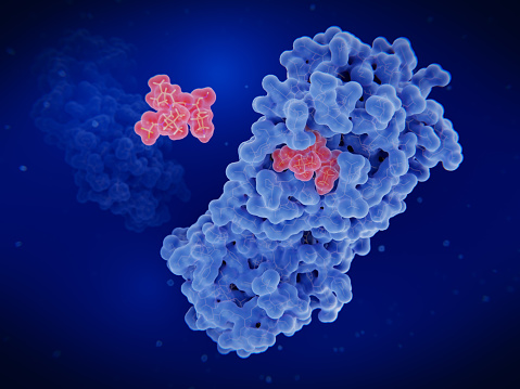
Free Images: "bestof:Porcine Mitochondrial IDH 1lwd.jpg Crystal Structure of Porcine Mitochondrial IDH enzyme in E coli From PDB 1LWD Created using Chimera Crystal structure of"
pattern-structure-crystal-structure-687736.jpg
pattern-structure-crystal-structure-687735.jpg
stone-quartz-mineral-481956.jpg
pattern-glass-patterns-structure-687737.jpg
crystal-rock-stone-natural-stones-844846.jpg
gem-grinding-ground-structure-pink-585082.jpg
crystal-mineral-quartz-rock-crystal-605641.jpg
purpurit-mineral-341399.jpg
sodalite-mineral-341397.jpg
purpurit-mineral-341396.jpg
mineral-iridescent-bismuth-81349.jpg
mineral-iridescent-bismuth-80056.jpg
snow-crystal-blue-cold-winter-ice-1174386.jpg
glazed-includes-mineral-iridescent-91860.jpg
glazed-includes-mineral-iridescent-91858.jpg
glazed-includes-mineral-iridescent-91859.jpg
glazed-includes-mineral-iridescent-91854.jpg
glazed-includes-mineral-iridescent-91855.jpg
glazed-includes-mineral-iridescent-91857.jpg
glazed-includes-mineral-iridescent-91853.jpg
crystal-mineral-iridescent-bismuth-91278.jpg
glazed-includes-mineral-iridescent-81348.jpg
crystal-mineral-iridescent-bismuth-81347.jpg
purpurit-sodalite-mineral-341395.jpg
glass-crystal-colorful-585631.jpg
ice-finnish-cold-frost-snow-1170573.jpg
rime-frost-pakkaskukka-1112815.jpg
frost-patterns-flower-ice-979757.jpg
val-st-lambert-belgium-crystal-142995.jpg
snow-snowflakes-winter-ice-crystal-1166061.jpg
abstract-glass-pattern-architecture-379799.jpg
snow-crystals-eiskristalle-625172.jpg
2ph1 pdb gallery nucleotide binding-protein-2 (MinD homolog).svg
Minocycline structure.svg
P450cycle.svg
Structure martensite.svg
Disloc alliage ordonne.svg
Structure FeAl B2.svg
Clindamycin skeletal.svg
Glissement facile distance mouvement.svg
Sr 2 Ru O 4 Layered Perovskite Structure.svg
alunite-crystal-structure-solid-875821.png
HCP crystal structure.svg
FCC crystal structure.svg
BCC crystal structure.svg
anhydrite-calcium-sulfate-crystal-876167.png
NaCl-estructura cristalina.svg
Cryst struct deficit.svg
ring-about-diamond-noble-valuable-449342.png
Pixantrone 2.svg
Mesitylgold.svg
Griseofulvin.svg
Phthalates.svg
Cinnamyl alcohol.svg
Inositol monochrome.svg
2-propanone.svg
Phytochelatin.svg
Uranium dioxide.svg
N-Ethyltryptamine.svg
1,2,4-Trioxane.svg
Fumagillin.svg
Amphotericin B structure.svg
Tetrabenazine structure.svg
Crizotinib structure.svg
Losartan structure.svg
Givinostat structure.svg
Triazolam structure.svg
Candoxatril-skeleton(2).svg
Semagacestat structure.svg
blocks-fractal-sponge-cube-square-655069.jpg
NaCl-Ionengitter.svg
18-crown-6.svg
Bullvalene.svg
Cephalosporin core structure.svg
Glibenclamide.svg
Formic acid.svg
Tc99 sestamibi 2D structure.svg
Epigallocatechin.svg
CP154526.svg
Spin glass by Zureks.svg
Trimedoxime bromide structure.svg
Beta-thujone.svg
Dexamethasone.svg
XAR File Structure.svg
Zearalenon.svg
CFT-Low Spin Splitting Diagram-Vector.svg
CFT-High Spin Splitting Diagram-Vector.svg
Cicutoxin.svg
Isocicutoxin.svg
Aethusin.svg
Sphingosine-1-phosphate.svg
Zearalanol.svg
Ochratoxin A.svg
Bullvalene Cope2.svg
Dinazafone.svg
TMC207 structure Andries et al.svg
CX157 structure.svg
Microzone.svg
Dabigatran structure.svg
Lanreotide.svg
Terms of Use   Search of the Day