Click Here for More Images from iStock- 15% off with coupon 15FREEIMAGES 
Cells of algae with chloroplast, Microscopic magnification
Diffuse large B-cell lymphoma, or DLBCL, is a cancer that starts in white blood cells called lymphocytes.
The Microscopic World. Onion epidermis with cells.
corn stem micrograph with dye
Epidermis. Electron microscope micrograph showing a keratinocyte of spinous layer. The epithelial cell has a polygonal shape, central nucleus with nucleolus, cytoplasm full of keratin filament bundles, and numerous dark desmosomes crossing the intercellular spaces.
Round, green chloroplasts in plant cells of anacharis or waterweed, Egeria densa. Chloroplasts carry chlorophyll which makes them green. These chlorplasts actually circulate around within each cell. Live specimen. Wet mount, 40X objective, transmitted brightfield illumination. Note - very shallow depth of field, chromatic aberration and uneven focus are inherent in light microscopy.
Host cells with spores (mold) are inside wood under the microscope for education.
3d Plant Cell structure isolated on white background
Micrograph of a moss leaf, Dicranum, with elongated cells, and an elongated midrib (costa) that may be the water conducting cells of the leaf, with polarization at 200x.
Myxoid liposarcoma.  Myxoid/round cell liposarcoma, or MRCLS, is one of several types of liposarcoma. Liposarcoma is a rare cancer that grows in the cells that store fat in the body. MRCLS usually grows in the arms and legs. These tumors grow slowly, and they can spread to other parts of the body
Plant tissue  under a microscope
plant Pumpkin ovary under light mircoscope with white background
Microscopic moss leafs
Particles, medical concept
Cell structure Hydrilla, view of the leaf surface showing plant cells under the microscope for classroom education.
Close up of cristal beads
Leiden leaf c.s. under microscope
Fatty liver disease (steatosis) is a common condition caused by having too much fat build up in your liver. A healthy liver contains a small amount of fat. It becomes a problem when fat reaches 5% to 10% of your liver's weight.
Microscopic examination of Stool showing Blastocystis hominis parasite.
Photomicrograph of stomata of leaf. Typical monocot distribution of stomata. Epidermis peeled from iris leaf. Live specimen. Wet mount, 40X objective, transmitted brightfield illumination.
Microalgae under microscopic view, green algae, cyanobacteria, phytoplankton, diatom, algae mix collage background
Squash preparation of  onion root tip cells stained to reveal chromosomes,  Bright field illumination, light microscopy.  This image was taken with the X40 objective lens.  Depth of focus is extremely low at this magnification.  Smaller apertures give greater depth of focus, but image quality deteriorates and resolution is greatly reduced.  The aperture in this image was optimised for contrast and resolution. Chromatic abberation is inevitable in light microscopy, but has been kept to a minimum in this image.
The study of plant tissues under the microscope in the laboratory.
Microscopic image of Microscopic image of nymphaea of aqustio stem
centropyxis aculeata (amoeboid organism) under the microscope - optical microscope x400 magnification
plant rape flower cross section under light mircoscope with white background
Water algae cells - microscope magnification
A close up of a computer CPU chip.
Plant cells have cell walls, constructed outside the cell membrane and composed of cellulose, hemicelluloses, and pectin.
Aerial view of natural shapes and textures on Dried Salt Lake. Aci Gol (Acı Göl) in Denizli, Turkey. Taken via drone.
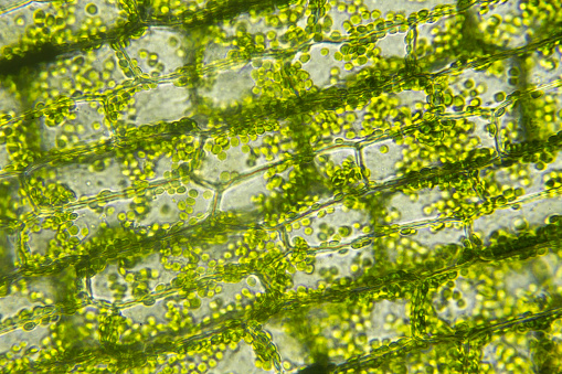
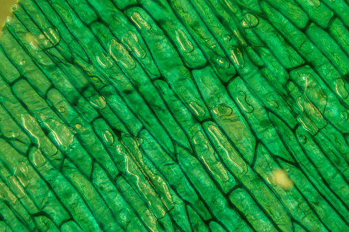
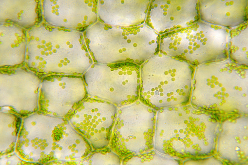
Free Images: "bestof:Plant cell wall nl.svg en plant cell wall Dutch nl celwand van plantencel File Plant cell wall diagram svg User LadyofHats 2013-01-25 Other versions/Plant cell"
Plant cell wall nl.svg
Plant cell wall diagram mk.svg
Plant cell wall diagram-uk.svg
Plasmolyse Pflanzenzelle.svg
Turgor pressure on plant cells diagram pl.svg
Turgor pressure on plant cells diagram-es.svg
Turgor pressure on plant cells diagram-nl.svg
Schema pflanzliche Zellwand.svg
Plant cell wall diagram-en.svg
Turgor pressure on plant cells diagram (zh-tw).svg
Turgor pressure on plant cells diagram.svg
Cell membrane detailed diagram nl.svg
Sciana komorkowa roslin diagram pl.svg
Plant cell structure svg labels.svg
Plant cell wall diagram gl.svg
Turgor pressure on plant cells diagram (zh-cn).svg
Plant cell structure-gl.svg
Average prokaryote cell- id.svg
Plant cell structure-en.svg
Plant cell structure svg 2.svg
Plant cell structure svg zh-hant.svg
Average prokaryote cell- nl.svg
Cellular tight junction keys.svg
Plant cell structure svg 2 sl.svg
Plant cell structure no text-2.svg
Plant cell structure svg-id.svg
Cell membrane detailed diagram en.svg
Endomembrane system diagram nl.svg
Turgor pressure on plant cells diagram-it.svg
Prokaryote cell diagram international.svg
Apoplast and symplast pathways Dutch txt.svg
Cell membrane detailed diagram blank.svg
Plant cell structure tr.svg
Plant cell structure svg-zh-cn.svg
Plant cell structure svg-es.svg
Plastids types en.svg
Plastids types zh.svg
Cell membrane detailed diagram id.svg
Animal cell structure id.svg
Turgor pressure on plant cells diagram-uk.svg
Complete neuron cell diagram tr.svg
Cell junction simplified it.svg
Average prokaryote cell- uk.svg
Average prokaryote cell- ru.svg
Diagram human cell nucleus oc.svg
Plant cell structure pt.svg
Plant cell structure cs.svg
Plant cell structure fr.svg
Plant cell structure svg hu.svg
Complete neuron cell diagram it.svg
Plant cell structure svg-de.svg
Plant cell structure svg pl.svg
Animal mitochondrion diagram it.svg
Plant cell structure svg uk.svg
Mature flower diagram NL.svg
Plastids types cs.svg
Plastids types es.svg
Chloroplast diagram (zh-cn).svg
Chloroplast diagram.svg
Diagram human cell nucleus tr.svg
Gap cell junction-en.svg
Animal cell cycle-en.svg
Cell membrane detailed diagram et.svg
Cell junction simplified en.svg
Animal cell structure gl.svg
Struttura della cellula vegetale.svg
Desmosome cell junction pl.svg
Cell membrane detailed diagram pl.svg
Diagram human cell nucleus sl.svg
Cell membrane detailed diagram ua.svg
Cell membrane detailed diagram de.svg
Endomembrane system diagram cs.svg
Cell junction simplified zh traditional.svg
Cell membrane detailed diagram lv.svg
Animal mitochondrion diagram unlabelled.svg
Animal cell structure ca.svg
Endomembrane system diagram en (edit).svg
Animal mitochondrion diagram en (edit).svg
Gap cell junction keys.svg
Animal cell structure sv.svg
Chloroplast diagram sr.svg
Average prokaryote cell- lt.svg
Adherens Junctions structural proteins-ja.svg
Endomembrane system diagram ka.svg
Prokaryote cell-ro.svg
Endomembrane system diagram en.svg
Flagellum base diagram keys.svg
Flagellum base diagram en.svg
Desmosome cell junction it.svg
Zelula prokarioto tipikoa.svg
Cell membrane detailed diagram es.svg
Cell membrane detailed diagram fr.svg
Scheme simple diffusion in cell membrane-pl.svg
Prokaryote cell diagram he.svg
Complete neuron cell diagram he.svg
Erythrozyten und Osmotischer Druck.svg
Animal cell structure en gl.svg
Complete neuron cell diagram numbered large numbers.svg
Prokaryote cell diagram cs.svg
Diagram human cell nucleus cs.svg
Terms of Use   Search of the Day