Click Here for More Images from iStock- 15% off with coupon 15FREEIMAGES 
Optical Coherence Tomography . ( OCT )
Cardiac amyloidosis is a condition where faulty proteins build up in your heart. You can inherit this condition, or it can develop on its own (usually later in life). As faulty proteins accumulate, your heart struggles to pump, ultimately leading to heart failure and death.
Cotton Candy on Vibrant Blue Background
Computational tomography image of the abdominal and pelvic regions.
Young female scientist loading a grid with an specimen on the sample holder of a transmission electron microscope
Mammogram
Hyaline cartilage, Elastic cartilage and Bone Human under the microscope in Lab.
Atmospheric photo of modern MRI scanning room in hospital. Non-invasive examination. Magnetic resonance imaging technology in specialized medical clinic.
Collection of  MRI heart or Cardiac MRI ( magnetic resonance imaging ) of heart  showing heart beating for detecting heart disease.
Carcinoid tumors are a type of slow-growing cancer that can arise in several places throughout your body. Carcinoid tumors, which are one subset of tumors called neuroendocrine tumors, usually begin in the digestive tract (stomach, appendix, small intestine, colon, rectum) or in the lungs. Site: Ovary
Prostate Cancer: Microscopic image of prostatic tissue, adenocarcinoma, malignant neoplasm, atypical epithelial cells.
Close-up of optometry slit lamp in a doctors office
Motor Neuron cells under the microscope
Dental Examination Equipment Close-up
ct scan step set of neck coronal view
Brush Grunge Background
Collection of CTA whole aorta with curve MPR technique showing abdominal aorta and left, right iliac artery showing aortic dissection .
Diffuse large B-cell lymphoma, or DLBCL, is a cancer that starts in white blood cells called lymphocytes.
Backgrounds of Bacteria and Mold under the microscope in Lab.
CT angiography of lower limb..artery occlusive disease
PAPS Smear study of a young women under microscopy showing Atrophic changes in uterus which is unusual at young age.
Clockwise, axial and sagittal T1 post contrast, then sagittal and axial T2 MRI of the pelvis with a large (11 cm) uterine fibroid with central necrosis, compressing the urinary bladder and rectum. Uterine fibroids are common benign tumors of the uterus that first appear during fertile years and grow with hormonal stimulation until menopause. They can be tiny and go unnoticed or grow as large as a full-term baby. In addition to expanding the waistline, fibroids can cause prolonged, heavy menstrual bleeding and pain, pelvic pain, and pressure on the bladder and rectum leading to difficulty with passing bowel movements and frequent urination. They can impair venous blood flow out of the legs, causing leg pain and heaviness. Most fibroids can be treated with a non-invasive procedure where the blood supply is cut off through a catheter threaded from the femoral artery into the uterine artery, termed uterine artery embolization or uterine fibroid embolization, performed by an interventional radiologist.
The right middle lobe bronchus is supplied from the right bronchial artery 3d illustration
Digital Dispersed corona virus on petri dish. Animation of multiplication of CORONA VIRUS COVID-19
CT scan of  Abdomen  sagittal view with injection contrast media with green color mode coronal view for diagnosis  abdominal diseases.
Plasmacytoid urothelial carcinoma (PUC) is a unique variant of invasive urothelial carcinoma characterized by tumor cells that exhibit a striking resemblance to plasma cells, and it is known to have an aggressive behavior.
Microscopic preparation, tissue plant
Surface photography of nature pattern Crystal shot for background.
Education Spinal cord, Nerve, Cerebellum, Cortex and Motor Neuron Human under the microscope in Lab.
Image taken with Flir T420 infra red digital camera. Each color represents different temperatures, as is shown on spectrum scale on right side of image.
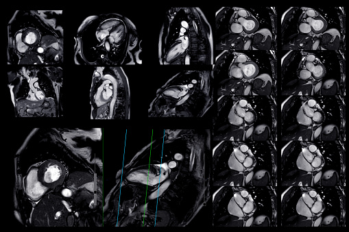
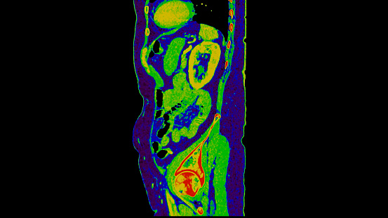
Free Images: "bestof:PerfAnimSag.gif Animated sequence from a reconstructed sagittal SPECT lung-perfusion study Generated by Kieran Maher using ImageJ 2006-10-22 KieranMaher"
PerfAnimSag.gif
CtAnimCor.gif
CtAnimSag.gif
VentAnim.gif
CtAnim.gif
PerfAnimCor.gif
PerfAnim.gif
Valves Of Heart Ultrasound.gif
Gyro.gif
CtOrthor.gif
MipParaPersp.gif
CtVRvascRed.gif
BModeLiverZoom - 2.png
HybridSpectCTScanner.png
ClutComp.jpg
OpacComp.jpg
NM16 44.jpg
HybridSpectCTScannerLabeled.png
SPECT CT fusion.jpg
Joint histograms for spatially shifted images.jpg
SegLungsCompos.gif
Histograms of pixel values in a bone scan and radiograph.gif
Histograms of pixel values in a bone scan and radiograph.jpg
SsdTransCompos.gif
SsdWFcompos.gif
ShadComp.jpg
Tomographic Imaging Labels.jpg
Scintillation Detector.gif
Histogram image against itself and spatially shifted.jpg
Low res CT scan for attenuation correction.jpg
Histograms of Bone Scan and Radiograph.jpg
VrParaPersp.gif
NM19 290.jpg
Nuclear Medicine 3-1.gif
Nuclear Medicine Ir1.jpg
Nuclear Medicine Ir6.jpg
Nuclear Medicine Ir9.jpg
Nuclear Medicine Ir10.jpg
Nuclear Medicine Ir11.jpg
Threshold LUT.gif
LiveImageB.jpg
TwoHandsGreyscale.jpg
Ct2dOrthoMPR.jpg
Nuclear Medicine Ir3.jpg
Nuclear Medicine Ir5.jpg
Nuclear Medicine Ir7.jpg
Nuclear Medicine Ir8.jpg
NM19 01.gif
UsMachTxPhoto.jpg
Ct2dMPRmax.jpg
Ct2dMPRmaxAngle.jpg
Ct2dOrthoMPRmax.jpg
Compton Effect.gif
Ssd2surfs.jpg
180HI Algorithm.png
180LI Algorithm.png
180MFI Algorithm.png
180MLI Algorithm.png
360LI Algorithm.png
ChestRadiographInverted.png
VrCropFrame.jpg
Nuclear Medicine 6-2.gif
Half Life.gif
NM13 34.jpg
NM19 124.jpg
ClutCompos.jpg
OpacCompos.jpg
ShadCompos.jpg
VrCrop.jpg
Radioactive Decay Law.gif
Image Fusion Interlacing Blending.jpg
ContEnhcCompos.jpg
NM13 33.jpg
Radioactive Decay Law Decay Constants.gif
Radioactive Decay Law Linear.gif
Nuclear Medicine 8-2a.gif
Matrix array multi-slice CT detector.jpg
Multi-slice Acquisition Using Adaptive Array.jpg
McqStatsNov06.jpg
Gamma Camera SPECT Filtered Back Projection Lungs.png
Gamma Camera SPECT Iterative Reconstruction Lungs.png
Bone scan different resolutions.jpg
EngCompos.jpg
BPNM13 8.jpg
NM16 42a.jpg
ZProject.jpg
GammaCameraTransmissionScanOfChest.png
3D MIP.gif
3D SSD.gif
CurvedMPR.jpg
BModeLiver.jpg
DualEnergyImageDemo.jpg
NM19 4.gif
ChestNL.jpg
NM19 121.jpg
NM19 122.jpg
T1t2PD.jpg
Dr1 emSpectrum.jpg
Dr1 eLevels.jpg
Dr1 eOrbits.jpg
Terms of Use   Search of the Day